Tinnitus Types: Subjective Tinnitus

The most common form: only you can hear the sound. What causes it, how doctors diagnose it, and what treatments are available.

  • Acoustic Neuroma and Tinnitus: Symptoms, Diagnosis, and What to Expect

    Acoustic Neuroma and Tinnitus: Symptoms, Diagnosis, and What to Expect

    One-Sided Ringing and a Diagnosis You Weren’t Expecting

    Hearing that a tumour might be the cause of ringing in your ear is frightening, even when a doctor reassures you it is benign. If you are in that position right now, you are dealing with something genuinely alarming, and that reaction makes complete sense. The good news is substantial: acoustic neuroma is non-cancerous, does not spread to other parts of the body, grows slowly (often over many years), and affects roughly 1 in 100,000 people per year. The medical term is vestibular schwannoma — acoustic neuroma is the older, more commonly used name, and both refer to the same thing.

    This article explains what acoustic neuroma is, why it causes one-sided tinnitus, how the diagnosis is reached, and — most importantly — what you can realistically expect regarding your tinnitus across the three main management paths.

    What Is Acoustic Neuroma and Why Does It Cause Tinnitus?

    Acoustic neuroma grows from Schwann cells on the vestibulocochlear nerve (cranial nerve VIII), the nerve responsible for both hearing and balance. As the tumour expands within the internal auditory canal, it compresses the cochlear branch of that nerve, disrupting the normal flow of auditory signals to the brain. The brain perceives this disruption as sound, which is the tinnitus you hear.

    Because the tumour sits on one side, this tinnitus is ipsilateral: it occurs in the same ear as the tumour. That one-sided, persistent quality is precisely what makes it clinically significant. Common tinnitus is usually bilateral or affects both ears at different times. When tinnitus is persistent and confined to a single ear, particularly when it is accompanied by hearing change on the same side, it is the defining red flag that warrants further investigation. Approximately 70% of people with acoustic neuroma have tinnitus at the point of diagnosis.

    Symptoms: What Acoustic Neuroma Feels Like

    Acoustic neuroma produces a recognisable pattern of symptoms, though their severity varies considerably depending on tumour size and how quickly it has grown.

    Progressive unilateral hearing loss is the most common symptom and is usually the first to appear. It tends to be gradual, affecting high frequencies first, and may be so slow that you attribute it to ageing or background noise. In around one in ten cases, hearing loss arrives suddenly rather than gradually, and sudden hearing loss in one ear is a medical urgency (more on this below).

    Tinnitus is present in roughly 70% of patients at diagnosis. It typically sounds like a persistent ringing, buzzing, or hissing, and occurs in the affected ear only. It may be constant or come and go. This ipsilateral quality — same ear as the hearing loss — is what separates acoustic neuroma tinnitus from the far more common bilateral tinnitus that affects millions of people without any structural cause.

    Vestibular symptoms — including dizziness, unsteadiness, or a sense of imbalance — are common because the tumour also affects the balance branch of cranial nerve VIII. Acute spinning vertigo (the room-spinning sensation of classic vertigo) is less typical; more often, people describe a general unsteadiness or feeling of being slightly off-balance.

    As the tumour grows larger, it may compress neighbouring structures, producing additional symptoms:

    • Facial numbness or tingling, from pressure on the trigeminal nerve (cranial nerve V)
    • Facial weakness, from involvement of the facial nerve (cranial nerve VII), which runs in close proximity
    • Headache or a feeling of pressure, which can develop if the tumour grows large enough to raise intracranial pressure

    Smaller tumours, which are increasingly found because of greater awareness and improved imaging, often produce only hearing loss and tinnitus, without any of these later-stage features.

    How Is Acoustic Neuroma Diagnosed?

    The diagnostic process follows a well-established sequence, and most small tumours are identified before they cause serious problems.

    Step 1: GP or ENT assessment. The process typically begins when you report persistent one-sided tinnitus, asymmetric hearing loss, or unexplained dizziness to your GP. Based on your symptom history, they will refer you for a hearing test or directly to an ENT specialist.

    Step 2: Audiogram. A formal hearing test (audiogram) is usually the first investigation. Acoustic neuroma typically produces asymmetric sensorineural hearing loss, meaning the nerve-based hearing loss is noticeably worse in one ear than the other. In the UK, NICE guidelines recommend MRI referral when there is an asymmetry of 15 dB or more at two adjacent frequencies (NICE NG98). An audiogram that shows this pattern is the key trigger for imaging.

    Step 3: MRI with gadolinium contrast. MRI is the gold standard for diagnosing acoustic neuroma. The gadolinium contrast agent makes even small tumours visible on the scan. CT scanning is not reliable for detecting small acoustic neuromas and may miss them entirely, which is why MRI is always preferred when this diagnosis is being considered.

    Two additional tests may be ordered to gather more information about nerve function:

    • Auditory brainstem response (ABR) testing assesses how efficiently the auditory nerve transmits signals to the brain
    • Electronystagmography (ENG) evaluates vestibular function and may reveal reduced response on the affected side

    Neither of these confirms the diagnosis on its own, but both can guide the clinical picture before or alongside MRI.

    The Three Management Options — and What They Mean for Your Tinnitus

    This is where acoustic neuroma management differs from what many patients expect, and where honest information matters most.

    There are three established approaches: watchful waiting (observation), microsurgery, and stereotactic radiosurgery. The 2024 Clinical Practice Guideline on vestibular schwannoma management confirms that none of these approaches consistently eliminates tinnitus, and that treatment decisions should be made through shared decision-making based on tumour size, growth rate, symptoms, and patient preference (Lassaletta et al., 2024). There is almost never a clinical reason to rush a decision.

    Watchful waiting (observation)

    For small or stable tumours, active monitoring with serial MRI scans every 6 to 12 months is a legitimate and commonly chosen path. The aim is to detect any significant growth before it becomes a problem, rather than to treat a tumour that may never progress meaningfully.

    From a tinnitus perspective, watchful waiting neither reliably worsens nor improves it. A systematic review comparing watchful waiting against stereotactic radiosurgery in 1,635 patients found no significant difference in tinnitus outcomes between the two groups (Vasconcellos et al., 2024). This is both reassuring and realistic: observation is not a passive acceptance of worsening symptoms, but it is not a tinnitus treatment either.

    Microsurgery

    Surgical removal aims to take out the tumour entirely. For many patients, particularly those with larger or growing tumours, it remains the most appropriate option.

    Regarding tinnitus, the evidence is clear and patients deserve to know it: surgery does not reliably eliminate tinnitus. A systematic review and meta-analysis of 13 studies involving 5,814 patients found no significant difference in tinnitus outcomes between microsurgery and stereotactic radiosurgery, and the authors concluded that “no definitive conclusions could be drawn favouring either treatment” (Ramkumar et al., 2025). A separate observational study of 450 surgical patients found that surgery can worsen pre-existing tinnitus, and can even trigger new-onset tinnitus in patients who had none beforehand (Geng et al., 2025). Patients with serviceable hearing before surgery faced higher odds of both worsened and new-onset tinnitus post-operatively.

    Hearing preservation is more likely when the tumour is smaller and detected early, which is another reason prompt investigation of one-sided symptoms matters.

    Stereotactic radiosurgery (e.g., Gamma Knife)

    Radiosurgery uses precisely targeted radiation to stop the tumour from growing; it does not remove the tumour. Most patients treated this way retain a stable but present tumour for the rest of their lives, without it causing further harm.

    Tinnitus outcomes after radiosurgery are similarly variable and unpredictable. A network meta-analysis across multiple treatment modalities suggested radiosurgery may offer a slight advantage over microsurgery for tinnitus improvement, though the certainty of evidence was rated low given that most included studies were observational rather than randomised (Huo et al., 2024). Radiosurgery’s main advantage is avoiding the operative risks of open surgery while still controlling tumour growth.

    The honest picture

    Across all three paths, the consistent finding is that tinnitus outcomes are unpredictable. Some people see improvement; others experience no change; a proportion find tinnitus worsens, particularly after surgery. What treatment does reliably accomplish is controlling the tumour, and for a benign growth that is not going to spread, that is the primary goal. Tinnitus management after diagnosis typically involves the same approaches used for tinnitus of other causes: counselling, sound therapy, and hearing rehabilitation where relevant.

    When to See a Doctor: Red Flags You Shouldn’t Ignore

    If you have tinnitus and are wondering whether it warrants medical attention, the following guidance is intended to help you decide clearly, without alarm, but without delay where delay matters.

    Persistent tinnitus in one ear only, particularly if it has lasted more than a few weeks and is accompanied by any hearing change on the same side, should prompt a visit to your GP to arrange an audiogram. Most one-sided tinnitus has far more common causes than acoustic neuroma, such as earwax, middle ear fluid, or noise exposure, but acoustic neuroma is the most important condition to exclude, which is why the investigation pathway exists.

    Sudden hearing loss in one ear is a medical urgency. If you wake up with significantly reduced hearing in one ear, or if hearing drops sharply over a few hours, seek same-day medical attention. Corticosteroid treatment for sudden sensorineural hearing loss should begin as soon as possible, ideally within the first two weeks; benefit has been reported up to six weeks from onset, but outcomes are better with earlier treatment (AAO-HNS 2019 CPG). Do not wait for a routine appointment.

    Tinnitus combined with dizziness, balance problems, or facial weakness or numbness warrants prompt ENT referral, as this combination suggests involvement of structures beyond the cochlear nerve alone.

    Acoustic neuroma affects roughly 1 in 100,000 people per year. The vast majority of one-sided tinnitus is not caused by a tumour. But the investigation, an audiogram followed by MRI if asymmetry is confirmed, is straightforward, and identifying a small acoustic neuroma early gives you and your clinical team the widest range of options.

    Key Takeaways

    Acoustic neuroma is a rare but important cause of one-sided tinnitus. It is benign, does not spread, and in most cases grows slowly enough that you and your doctors have real time to consider options carefully.

    The key red flag is persistent tinnitus in one ear, especially when combined with hearing loss on the same side. That combination warrants an audiogram and, if asymmetry is confirmed, an MRI.

    If you receive a diagnosis, the most important thing to understand upfront is that none of the three management options, whether observation, surgery, or radiosurgery, reliably eliminates tinnitus. Knowing this from the start allows you to set realistic expectations and focus treatment decisions on what they do achieve: controlling the tumour. Diagnosis is not a crisis. Most people with acoustic neuroma lead full, active lives.

  • Left Ear Ringing: Causes, Red Flags, and When to See a Doctor

    Left Ear Ringing: Causes, Red Flags, and When to See a Doctor

    That Ringing in Your Left Ear: Why It Feels Different

    Noticing that only one ear is ringing — particularly late at night when everything is quiet — can be unsettling in a way that symmetrical sounds are not. There is something about the one-sidedness that makes it feel pointed, deliberate, worth worrying about. You are right to pay attention to it. In most cases, left-ear ringing has a benign explanation: earwax, a recent cold, or noise exposure. But the asymmetry does matter clinically, and this article explains why, which symptoms should prompt urgent care, and what to expect if you see a doctor.

    What Does It Mean When Only Your Left Ear Is Ringing?

    Ringing in only one ear — called unilateral tinnitus — is clinically significant because it warrants investigation to rule out serious causes, including a benign tumour on the auditory nerve known as acoustic neuroma; however, the most common causes are benign, such as earwax build-up or noise exposure, and acoustic neuroma accounts for only about 0.08% of cases where tinnitus is the sole symptom (Javed et al., 2023). One-sided tinnitus is less common than bilateral tinnitus and draws medical attention for a specific reason: the localisation suggests a structural or vascular issue in or near that ear, rather than a systemic process affecting both ears. The vast majority of people investigated for unexplained unilateral tinnitus are reassured after a clear audiogram and, where needed, a clear MRI.

    Common Causes of Left Ear Ringing

    Most cases of one-sided ringing come down to something localised and treatable. Here are the causes doctors consider first.

    Earwax impaction is the most common and most straightforward cause. When wax blocks the left ear canal, it raises pressure within the ear, which can produce low-pitched, one-sided ringing. The sound typically resolves after the wax is removed by a nurse or GP.

    Noise-induced hearing loss can be asymmetric when noise exposure is asymmetric. Musicians who sit with one ear facing amplifiers, drivers who spend hours with a window open on one side, or people who use a single earbud frequently in the same ear can develop tinnitus in just one ear. Occupational noise exposure — a drilling machine to one side, for example — follows the same logic.

    Ear infections and fluid are common triggers. Otitis media (middle ear infection) or otitis externa (outer ear canal infection) affecting only the left ear will produce one-sided symptoms including ringing, pain, and muffled hearing. Both are usually self-limiting or respond to appropriate treatment.

    Eustachian tube dysfunction explains a significant proportion of post-cold ear ringing. The Eustachian tube connects the middle ear to the back of the throat. After a sinus infection or upper respiratory virus, one tube can remain blocked for days to weeks, producing one-sided pressure, fullness, and intermittent ringing. Most cases resolve as the inflammation clears.

    Ototoxic medications — drugs that can affect hearing or balance — include high-dose aspirin and salicylates, certain aminoglycoside antibiotics, loop diuretics such as furosemide, and some chemotherapy agents. These usually cause bilateral effects, but they can present asymmetrically. If you recently started a new medication and noticed the ringing, mention it to your prescribing doctor.

    TMJ (temporomandibular joint) dysfunction is an underrecognised cause. The jaw joint sits close to the ear canal, and problems with jaw alignment, grinding, or clenching can produce one-sided ringing or clicking sensations that are often worse on waking or after eating. A dentist or maxillofacial specialist can assess this.

    The reassuring common thread across most of these causes is that the tinnitus typically improves or resolves once the underlying issue is treated.

    Conditions That Can Cause One-Sided Tinnitus — and Why Laterality Matters

    When a doctor sees a patient with one-sided tinnitus, their first job is to look for a localised cause — because unilateral tinnitus is a clinical red-flag category in its own right. Clinical guidelines from both the American Academy of Family Physicians and NICE recommend assessment for all patients with unexplained unilateral tinnitus (American Family Physician (2021); NICE (2020)). Here are the conditions that explain why.

    Ménière’s disease classically begins in one ear and produces a distinctive triad: low-frequency roaring tinnitus, episodic vertigo lasting minutes to hours, and fluctuating hearing loss. Ear fullness is also common. The condition tends to start unilaterally, though over years it can involve the other ear in some patients. There is no cure, but treatments can reduce the frequency and severity of episodes.

    Acoustic neuroma (vestibular schwannoma) is the condition many people fear when they notice one-sided ringing. It is a benign, slow-growing tumour on the vestibular nerve. Typical presentation includes progressive one-sided hearing loss, persistent unilateral tinnitus, and sometimes balance disturbance. It is genuinely rare: a systematic review of 1,394 patients who had MRI specifically for unilateral tinnitus without any hearing loss found a vestibular schwannoma rate of just 0.08% (Javed et al., 2023). The risk rises to around 2.22% when asymmetric hearing loss is also present (Abbas et al., 2018). Red-flag features that suggest a larger tumour and escalate urgency include facial weakness or numbness, balance problems, and headache (Foley et al., 2017). The rarity of the diagnosis is worth holding onto — but the reason doctors investigate is precisely because catching it early makes management more straightforward.

    Sudden sensorineural hearing loss (SSHL) deserves its own attention because the timing of treatment affects the outcome. If the left-ear ringing came on abruptly — within hours — and is accompanied by muffled or reduced hearing, this is a medical urgency. Steroids are used as soon as possible for the best effect; treatment delayed beyond two to four weeks is less likely to reverse permanent hearing loss (NIDCD / NIH (2023)). Approximately 85% of those who receive prompt treatment experience partial or full hearing recovery (NIDCD / NIH (2023)). Do not wait and see.

    Pulsatile tinnitus is a distinct type of one-sided ringing that pulses in time with your heartbeat rather than producing a constant tone. In contrast to the steady hiss or ring of typical tinnitus, pulsatile tinnitus has an identifiable vascular cause in the majority of cases (Herraets et al., 2017). Causes include arteriovenous malformations, high blood pressure, vascular tumours, and abnormal blood flow near the ear. One-sided pulsatile tinnitus always warrants investigation.

    Red Flags: When Left-Ear Ringing Needs Prompt Medical Attention

    Most cases of left-ear ringing are not emergencies. But specific patterns change that calculation. Here is a practical framework.

    Seek same-day or emergency care

    • Sudden ringing in the left ear paired with sudden muffled, reduced, or lost hearing. This is a possible sudden sensorineural hearing loss — treatment needs to start as soon as possible. Do not wait for a routine appointment.
    • Pulsatile (heartbeat-matching) ringing in one ear, especially with headache, vision changes, or neck pain. This may indicate a vascular cause requiring urgent imaging.
    • One-sided tinnitus with facial weakness, facial numbness, or sudden loss of balance. These features are associated with larger acoustic neuromas or neurological causes and require same-day assessment (Foley et al., 2017).

    See a GP or audiologist within one to two weeks

    • New left-ear ringing with no obvious cause — no recent loud noise, no cold, no wax build-up.
    • Left-ear ringing with gradual hearing loss or muffling on that side.
    • Ringing with recurring dizziness or a sense of ear fullness.
    • Left-ear ringing that began after a head or neck injury.

    For this group, AAFP guidelines recommend prompt audiometry and, where asymmetric hearing loss is confirmed or the cause remains unexplained, MRI of the internal auditory canals (American Family Physician (2021)).

    Monitor and book a routine appointment if persistent

    • Ringing that appeared after a cold or ear infection and is gradually improving.
    • Brief ringing after loud noise exposure that fades within a few hours.
    • Mild, intermittent ringing with no other symptoms.

    Even in this lower-urgency group, tinnitus that persists beyond a few weeks without an obvious trigger is worth discussing with a GP.

    All unexplained unilateral tinnitus — even without hearing loss or dizziness — warrants a GP visit to arrange a hearing test and, where clinically indicated, imaging. NICE (2020) recommends referral via local pathway for persistent unilateral tinnitus.

    What to Expect at the Doctor: Diagnosis and Next Steps

    If you go to your GP or audiologist with one-sided tinnitus, the appointment will typically follow a clear pathway — and knowing what to expect can make the visit feel less daunting.

    History and examination. Your doctor will ask when the ringing started, whether it is constant or intermittent, whether it pulses in time with your heartbeat, and whether you have noticed any change in your hearing. They will ask about recent noise exposure, medications, ear infections, jaw problems, and any associated dizziness or neurological symptoms.

    Audiogram. A comprehensive hearing test is the standard first investigation. It maps your hearing across a range of frequencies and identifies whether there is asymmetric sensorineural hearing loss — a finding that significantly raises the priority for imaging.

    MRI referral. If the audiogram shows asymmetric hearing loss, or if the tinnitus is unexplained and persistent, an MRI of the internal auditory canals is standard practice to exclude acoustic neuroma. AAFP guidelines explicitly mandate this for unilateral tinnitus associated with asymmetric hearing loss or where no cause is found (American Family Physician (2021)).

    Onward referral. Depending on findings, you may be referred to an ENT specialist or an audiology service for further management. Most people reach this point only to receive reassurance — a clear audiogram and, if required, a clear MRI is the most common outcome.

    Many people who see a doctor for one-sided tinnitus describe the audiology appointment as the moment their anxiety eased. Hearing a professional say the audiogram looks normal — and knowing they have been properly assessed — tends to shift the experience of the sound itself. Reassurance backed by a test is more useful than reassurance backed by nothing.

    Key Takeaways

    • Ringing in just your left ear (unilateral tinnitus) is clinically more significant than bilateral tinnitus. It always merits investigation because a localised cause needs to be found or excluded.
    • The most common causes are benign: earwax, ear infections, Eustachian tube dysfunction, and asymmetric noise exposure. Most respond to treating the underlying issue.
    • Serious causes such as acoustic neuroma are rare. In patients with unilateral tinnitus alone and no hearing loss, the detection rate is around 0.08% (Javed et al., 2023). Risk rises with asymmetric hearing loss — which is exactly why an audiogram is the right first step.
    • Pulsatile one-sided tinnitus and sudden-onset ringing with hearing loss are urgent. Seek care as soon as possible — delays beyond two to four weeks reduce the chance of recovery from sudden hearing loss.
    • A routine audiogram is usually the first diagnostic step, and most people are reassured after it.

    Left-ear ringing is rarely an emergency — but knowing which patterns require prompt care and which are safe to watch gives you something far more useful than worry: a clear plan for what to do next.

  • Spiritual Meaning of Ear Ringing: Left Ear, Right Ear, and Both

    Spiritual Meaning of Ear Ringing: Left Ear, Right Ear, and Both

    Why Does My Ear Keep Ringing? The Meaning People Search For

    A sudden ring in one ear — especially in a quiet room late at night — tends to stop you in your tracks. It’s unexplained, a little unsettling, and when it keeps coming back, the question of what it means feels entirely natural. Millions of people search for a spiritual or symbolic explanation, and this article covers what different cultural traditions say. It also explains what medicine and science actually know, including why which ear is ringing can genuinely matter for your health.

    What Ear Ringing Actually Is

    Ear ringing is a medical phenomenon called tinnitus: the perception of sound — ringing, buzzing, hissing, or humming — with no external source. Globally, about 14.4% of adults experience it at some point, affecting over 740 million people worldwide (Jarach et al. (2022)). Spiritually, many traditions assign meaning to which ear is ringing, but these beliefs have no scientific foundation. What does matter medically is whether the ringing is in one ear or both, how long it lasts, and whether it comes with other symptoms like hearing loss or dizziness. Those factors can point to causes ranging from noise exposure to inner ear disorders that deserve professional attention.

    What Different Cultures and Spiritual Traditions Believe

    Before modern medicine had an explanation for tinnitus, cultures worldwide filled that gap with meaning. The human instinct to interpret an invisible, intrusive sensation as a signal from beyond is ancient — and it shows up across remarkably different traditions.

    Western folk belief is perhaps the most familiar: ringing in the right ear means someone is speaking well of you, while ringing in the left ear means you are the subject of gossip or criticism. This is documented as far back as Pliny the Elder in Naturalis Historia (~77 AD), where he noted ear-ringing omens among Roman customs.

    New Age and metaphysical traditions often assign a more elaborate framework. The left ear is said to receive inward, intuitive messages — sometimes interpreted as your own spiritual growth or energy shifts. The right ear is associated with external communications from spirit guides or higher realms. A high-pitched tone in either ear is sometimes read as a sign of spiritual awakening.

    Hindu traditions connect the ears to the nadi energy channels and to the primordial sound of Om. In this framework, ear ringing may be understood as a spiritual message or a sign of heightened awareness along the chakra system.

    Chinese folk traditions add another layer, with interpretations tied to the time of day when the ringing occurs — specific hours are said to indicate different types of messages or events.

    These are genuinely interesting traditions that reflect how humans across history have tried to make sense of an unsettling, invisible symptom. One thing they all share: they contradict each other. In some systems, left-ear ringing is a warning; in others, it’s a blessing. Right-ear ringing is positive in one tradition and neutral in another. That inconsistency doesn’t make these traditions less meaningful to the people who hold them — but it does suggest they tell us more about human meaning-making than about the physiology of the ear.

    What the Left Ear, Right Ear, and Both Ears Mean Medically

    From a medical standpoint, which ear is ringing does carry significance — just not for spiritual reasons. The significance is anatomical and clinical.

    Left-ear tinnitus may be slightly more common than right-ear tinnitus, at least in women. Some research suggests a left-ear predominance for inner ear conditions including tinnitus and Ménière’s disease, possibly reflecting greater cochlear vulnerability on the left side in female patients, perhaps linked to hormonal differences (Reiss & Reiss (2014)). This is a single observational study and should not be overstated, but it illustrates that laterality has a real physiological dimension — one that folklore assigned to gossip and spirit guides.

    Unilateral tinnitus (ringing in one ear only, either side) is the pattern that gets clinicians’ attention. A meta-analysis of 1,394 patients found that the risk of vestibular schwannoma (acoustic neuroma) in people with isolated unilateral tinnitus and no hearing loss is low — around 0.08% (Javed et al. (2023)). So persistent one-sided ringing is not cause for panic. When unilateral tinnitus is combined with asymmetric hearing loss, however, that risk picture changes, and investigation is warranted. Among patients diagnosed with acoustic neuroma, unilateral tinnitus is a presenting symptom in about 6.3% of cases (Foley et al. (2017)) — less common than hearing loss, but a genuine signal. UK clinical guidelines specify that persistent unilateral tinnitus warrants routine ENT referral, and tinnitus accompanied by sudden hearing loss warrants urgent assessment within 24 hours (NICE (2020)).

    Bilateral tinnitus (ringing in both ears) is more commonly linked to noise-induced or age-related hearing loss. It’s also more prevalent overall — most people who develop chronic tinnitus report it in both ears or describe it as centred in the head.

    Pulsatile tinnitus — a rhythmic, heartbeat-like sound rather than a constant tone — is a distinct type altogether. It typically has a vascular cause, and Mayo Clinic guidance recommends same-day or next-day consultation for pulsatile tinnitus or tinnitus with sudden hearing loss (Mayo (2024)). NICE guidelines call for immediate referral for sudden-onset pulsatile tinnitus (NICE (2020)).

    The bottom line: the ‘meaning’ of which ear is ringing lies in anatomy and pathology, not metaphysics. And for most people, it will turn out to mean nothing serious at all — but some patterns are worth a medical conversation.

    When Ear Ringing Is Just Ear Ringing — and When It Isn’t

    Brief, spontaneous ear ringing — a tone that appears for a few seconds and fades — is extremely common and almost always benign. It can follow noise exposure, happen in a very quiet room, or occur for no identifiable reason. This kind of transient ringing is part of normal auditory experience for most people.

    Persistent tinnitus is different. When ringing lasts more than a few days without a clear trigger, a visit to a GP or audiologist is a reasonable step. There’s no need for alarm, but there’s also no reason to ignore it.

    Certain patterns should prompt faster action:

    • Sudden onset of one-sided ringing with no obvious cause
    • Tinnitus with hearing loss — especially sudden or one-sided hearing loss
    • Tinnitus with vertigo or dizziness
    • Pulsatile tinnitus (rhythmic, in time with your heartbeat)
    • Tinnitus following head or neck trauma

    A practical guide: if ringing persists beyond 48 hours without a clear explanation like a loud concert, get it checked. Most causes will be straightforward, but some — Ménière’s disease, sudden sensorineural hearing loss, acoustic neuroma — are time-sensitive to treat or to rule out.

    The American Tinnitus Association estimates that around 2 million Americans find tinnitus debilitating (American (2024)). Many of those cases might have benefited from earlier evaluation. Seeking help isn’t overreacting — it’s the sensible response to a symptom your body is persistently signalling.

    If your ear ringing started suddenly, affects only one ear, is accompanied by hearing loss or dizziness, or sounds like a pulse, see a doctor promptly — ideally within 24 hours. Don’t wait to see whether it resolves on its own.

    Key Takeaways

    • Ear ringing is a medical symptom called tinnitus, with well-understood neurological and physiological causes. It affects roughly 14% of adults globally (Jarach et al. (2022)). There is no scientific evidence for a spiritual meaning.
    • Many cultures across history have assigned meaning to left vs. right ear ringing — from Roman omens to New Age energy frameworks. These are genuinely interesting traditions, but they contradict each other across cultures, which tells you something about their nature.
    • Which ear rings does matter medically. Unilateral (one-sided) tinnitus is a more significant clinical finding than bilateral tinnitus and warrants an ENT assessment, particularly when it comes with hearing changes or dizziness.
    • Red flags that mean see a doctor, not search for omens: sudden onset, one-sided ringing, ringing with hearing loss or vertigo, pulsatile ringing, or ringing after head trauma.
    • Brief, occasional ringing is common and usually harmless. Ringing that persists beyond 48 hours deserves professional evaluation.

    If you are searching for what your ear ringing means, that concern is worth taking seriously — just take it to a doctor rather than a horoscope. Most causes are benign, and the ones that aren’t respond better to early attention.

  • COVID and Tinnitus: What the Research Says About Onset and Recovery

    COVID and Tinnitus: What the Research Says About Onset and Recovery

    Why Is My Ear Ringing After COVID?

    If you’ve recovered from COVID-19 and now have a ringing, buzzing, or humming in your ears that wasn’t there before, it’s natural to feel alarmed. You might be wondering whether this is connected to your illness, whether it will go away, and whether you need to see a doctor. These are the right questions to ask, and there are real answers.

    This article covers how common tinnitus after COVID actually is, why the infection can affect your hearing, and what the evidence says about recovery. The short version: COVID tinnitus is a documented, recognised phenomenon. Whether it resolves depends in part on how severe it is at onset, and that distinction matters for what you do next.

    Can COVID-19 Cause Tinnitus?

    Yes. COVID-19 is associated with new-onset tinnitus and the worsening of pre-existing tinnitus. Depending on the study and the population examined, somewhere between roughly 5% and 28% of people who have had COVID-19 report tinnitus afterwards.

    The range is wide because it reflects genuine differences in study design. A 2022 meta-analysis of 12 studies found a pooled tinnitus rate of around 4.5% across largely hospital-based acute-phase cohorts (Jafari et al., 2022). A larger cross-sectional survey of 1,331 post-COVID respondents found a prevalence of 27.9% (Mao et al., 2024). A 2026 meta-analysis of cohort studies using physician-diagnosed outcomes found no statistically significant pooled association overall (Liu et al., 2026), which shows how much the answer depends on who is studied and how tinnitus is measured.

    COVID-19 can trigger new-onset tinnitus in a meaningful proportion of survivors. Estimates vary widely across studies — from around 5% to 28% — depending on whether researchers studied hospitalised patients, mild-case survivors, or long-COVID clinic populations. The figure is real, even if the exact number is uncertain.

    What is consistent across studies is that the association is real and that it affects people across the spectrum of COVID severity, not just those who were seriously ill. Worsening of pre-existing tinnitus is also well-documented.

    When Does COVID Tinnitus Start — and Why Does Timing Matter?

    Not everyone who develops tinnitus after COVID notices it at the same point in their illness. Research points to three distinct onset windows, and understanding which applies to you can help clarify what is likely driving it.

    During the acute illness phase. Some people notice tinnitus while they are still actively sick — during the first one to two weeks of infection. This most likely reflects direct cochlear involvement: inflammation, reduced blood flow, or early viral effects on the inner ear during the height of the immune response.

    During treatment. A subset of cases appear to begin during COVID treatment rather than from the infection itself. Corticosteroids, sometimes prescribed for COVID, are among the medications that can independently affect tinnitus perception. Separating drug effects from viral effects in this window is genuinely difficult, and the research doesn’t fully resolve it.

    After recovery — delayed onset. Some people develop tinnitus days or weeks after they have otherwise recovered. One audiometric study found that tinnitus onset averaged around 30 days after the initial COVID symptoms. This delayed pattern may reflect a different underlying process: post-inflammatory changes in the central auditory system, or ongoing immune activation rather than the direct cochlear effects more likely in the acute phase.

    The timing matters clinically because it shapes how you understand the likely cause. Tinnitus appearing during acute illness suggests peripheral (inner ear) involvement. Tinnitus appearing weeks after recovery, without any other hearing change, is more likely to involve central auditory pathways — a distinction that affects how the condition is managed.

    Why Does COVID Affect Your Hearing? The Biology in Plain Language

    Your cochlea — the spiral-shaped structure in your inner ear that converts sound into nerve signals — contains cells that carry a protein on their surface called ACE2. This is the same receptor that SARS-CoV-2 uses to enter cells throughout the body. Animal studies have confirmed that ACE2, along with related proteins that help the virus enter cells, is present in cochlear hair cells, the stria vascularis, and the spiral ganglion (Uranaka et al., 2021). This establishes the biological plausibility that the virus can, in principle, directly affect the inner ear.

    Here is the chain of events researchers believe may occur:

    1. Viral or inflammatory damage to cochlear hair cells. Hair cells are the sensory cells that detect sound vibrations. They do not regenerate once lost. If the virus or the immune response triggered by it damages these cells, the cochlea sends fewer signals to the brain.

    2. The brain compensates by turning up its internal volume. When the brain receives less input from the ear, it tends to amplify its own activity to compensate. This process — called central gain upregulation — can produce phantom sounds that feel just as real as external noise. That is tinnitus.

    3. Auditory pathway involvement beyond the cochlea. Objective audiometric testing of long-COVID patients found significantly prolonged signal transmission times through the brainstem auditory pathway, suggesting that nerve damage extends beyond the inner ear itself (Dorobisz et al., 2023).

    4. Mechanical causes from the upper airway. Eustachian tube dysfunction — common during and after any upper respiratory infection — can cause ear fullness and muffled hearing that temporarily triggers or worsens tinnitus through a simpler mechanical route, without any cochlear damage at all.

    No single mechanism has been confirmed as the primary cause of COVID-related tinnitus, and it likely varies between individuals. Anxiety and poor sleep — both common during and after COVID illness — can independently intensify tinnitus perception regardless of the underlying cause. Some COVID medications may also play a role.

    If your tinnitus started during COVID or shortly after, you are not imagining it and you are not alone. The biological pathways described above are plausible and supported by evidence, even though researchers are still working out exactly which pathway dominates in different cases.

    Will COVID Tinnitus Go Away? What the Research Actually Shows

    This is the question most people searching this topic most want answered. The honest answer is: it depends on how severe it is.

    The most detailed evidence on this comes from Mao et al. (2024), whose survey of 1,331 post-COVID respondents found a clear severity gradient in outcomes. Mild (Grade I) tinnitus had notably higher rates of spontaneous resolution. Severe tinnitus — classified as Grade IV — had low spontaneous resolution rates and a strong association with long-term hearing loss and anxiety disorders. Grade IV was also the most common severity grade reported, representing 33.2% of all tinnitus cases in the survey.

    This matters for what you do next. If your tinnitus is mild and fading, watchful waiting with good sleep and stress management is reasonable. If it is severe, intrusive, or has not improved after several weeks, waiting longer is unlikely to help and may delay treatment that could.

    A smaller audiometric study of long-COVID patients with hearing complaints found that, at around 259 days post-infection, 7 out of 21 patients who had presented with tinnitus showed full recovery; 14 had only partial recovery or none at all (Dorobisz et al., 2023). This is a small sample and cannot be generalised widely, but it is consistent with the pattern from Mao et al.: a substantial proportion of cases do not resolve without support.

    Hospitalisation history is also a relevant predictor. Research has found that patients who were hospitalised during their COVID illness tend to have worse tinnitus outcomes than those with milder acute illness, with severity correlating significantly with hospitalisation status.

    Severe or persistent tinnitus after COVID is not likely to resolve on its own without support. If your tinnitus has lasted more than a few weeks after your COVID illness and is significantly affecting your daily life or sleep, seek an audiological evaluation rather than waiting indefinitely.

    Importantly, this does not mean severe cases are untreatable. Standard tinnitus management approaches — including cognitive behavioural therapy, sound therapy, and audiological support — can reduce distress and improve function even when spontaneous resolution does not occur. Severity at onset is the best available predictor of whether the tinnitus will resolve on its own; it does not determine whether you can get better with the right support.

    COVID Tinnitus vs. Long COVID Tinnitus: Is There a Difference?

    You may have heard the term “long COVID” and wondered whether it applies to you. Under NICE guidance, long COVID (formally called post-COVID-19 syndrome) is defined as symptoms that develop during or after COVID infection, persist for more than 12 weeks, and cannot be explained by another diagnosis. Tinnitus is explicitly listed as a recognised ENT symptom of long COVID under these guidelines (NICE/SIGN/RCGP, 2024).

    The clinical categories break down like this:

    • Acute COVID: symptoms lasting up to 4 weeks
    • Ongoing symptomatic COVID: symptoms lasting 4 to 12 weeks
    • Post-COVID-19 syndrome (long COVID): symptoms lasting 12 weeks or more

    If your tinnitus has persisted beyond three months after your COVID illness, it qualifies as a recognised long COVID symptom — which matters because it entitles you to appropriate clinical assessment and support rather than being dismissed as something unrelated.

    Long COVID tinnitus may involve a somewhat different biological dynamic than tinnitus that resolves in the acute phase. Persistent systemic inflammation, central sensitisation, and possible autoimmune mechanisms are all proposed contributors. A 2025 narrative review found that approximately 1 in 5 long-COVID patients reports tinnitus (Guntinas-Lichius et al., 2025). Self-reported rates in long-COVID populations are often higher.

    None of this means long COVID tinnitus is untreatable. It does mean it is less likely to resolve without some form of structured support, and more likely to respond well if you seek it.

    What Can You Do If You Have COVID Tinnitus?

    There is no treatment that specifically targets COVID tinnitus as a separate category — the same evidence-based approaches used for tinnitus from any cause apply here (Guntinas-Lichius et al., 2025). The practical steps below are grounded in what the research supports.

    See a GP or ENT if tinnitus has lasted more than a few weeks. Do not wait indefinitely. Ask for a referral for audiological evaluation to check for underlying hearing loss, which may accompany the tinnitus and is worth detecting early.

    Manage the things that make tinnitus louder. Anxiety, poor sleep, and sustained stress are known amplifiers of tinnitus perception — and all three are common during post-COVID recovery. Improving sleep quality and managing anxiety are not just general wellness advice; they have a direct effect on how tinnitus is perceived.

    Standard tinnitus therapies apply. Cognitive behavioural therapy for tinnitus has strong evidence for reducing tinnitus-related distress. Sound therapy and audiological counselling are also established options. Your GP or an audiologist can help you access these.

    If you had tinnitus before COVID and it has worsened, this is also documented and worth raising with a clinician. A small controlled study found that COVID infection itself — not just pandemic stress — significantly worsened tinnitus severity and quality of life in people with pre-existing tinnitus, even without changes in hearing thresholds (Aydogan et al., 2025). You are not imagining a deterioration.

    What This Means for You

    If you came to this article worried about a new ringing in your ears after COVID, here is what the evidence actually shows.

    First, COVID tinnitus is real. It is documented across multiple large studies, officially recognised in clinical guidelines, and not imagined or exaggerated. You are not the only person dealing with this.

    Second, the prognosis is genuinely variable, and severity at onset is the most useful guide. Mild tinnitus that appeared during or shortly after COVID illness often improves over weeks to months. Severe tinnitus — particularly the intrusive, high-grade kind that affects sleep and daily functioning — is less likely to resolve on its own and more likely to need active management. Waiting without seeking help is rarely the right approach if tinnitus is severe or has persisted for weeks.

    Third, this is not an untreatable condition. There is no special “COVID tinnitus treatment,” but there are effective management approaches that work for post-COVID cases just as they do for other forms of tinnitus. Getting an audiological assessment is the right starting point — not because something is necessarily seriously wrong, but because knowing what you are dealing with puts you in a better position to manage it.

    The uncertainty can be hard to sit with. But understanding what is happening, and knowing when to seek support, is a meaningful first step.

  • Clogged Ear vs. Tinnitus: How to Tell the Difference and What Helps

    Clogged Ear vs. Tinnitus: How to Tell the Difference and What Helps

    That Stuffy, Ringing Ear: Why It’s Hard to Know What’s Going On

    You know the feeling: an ear that won’t pop after a flight, a low hum that appeared during a cold and hasn’t left, or a pressure that makes sounds feel muffled and distant. When both symptoms hit at once — a blocked sensation and a ringing or buzzing that won’t quit — it’s natural to wonder whether something is seriously wrong. The good news is that most of the time, both symptoms share one straightforward cause, and fixing that cause fixes both. But knowing when that’s true, and when it isn’t, is exactly what this article is for.

    Clogged Ear vs. Tinnitus: What’s the Difference?

    A clogged ear and tinnitus often occur together, but they are not the same thing: a clogged ear is a physical blockage or pressure imbalance in the outer or middle ear, while tinnitus is the brain’s perception of sound — ringing, buzzing, hissing — without an external source. When a blockage is the cause of the ringing, treating the blockage usually makes the tinnitus resolve too. The key distinction is whether the ringing comes from the blockage or exists independently of it.

    Why a Clogged Ear Can Cause Ringing

    The ear works as a mechanical system. Sound waves travel down the ear canal, vibrate the eardrum, pass through the three tiny bones of the middle ear, and reach the cochlea — the snail-shaped organ in the inner ear that converts those vibrations into electrical signals your brain interprets as sound.

    When something interrupts that pathway, the cochlea receives a different acoustic signal than it expects. A build-up of earwax, a pool of fluid behind the eardrum, or a blocked Eustachian tube all reduce or distort the sound that arrives at the cochlea. In response, the cochlea or the auditory pathways further up the chain can generate phantom signals — sounds that aren’t there. This is called conductive tinnitus, and the key thing to know about it is that it is typically temporary.

    The three most common causes are:

    • Cerumen (earwax) impaction: Wax that has built up and hardened in the ear canal physically blocks sound transmission. Tinnitus is a recognised symptom of cerumen impaction, alongside hearing loss and a sensation of pressure (Michaudet & Malaty, 2018).
    • Eustachian tube dysfunction: The tube that connects your middle ear to the back of your throat — and keeps air pressure equalised — can become blocked after a cold, hay fever, or a change in altitude. The resulting pressure imbalance creates that familiar underwater or muffled feeling, and often a low-frequency hum.
    • Middle ear fluid (otitis media): Fluid trapped behind the eardrum after an ear infection acts as a dampener on sound conduction, and can produce both muffled hearing and tinnitus until it drains.

    All three causes are among the most reversible. Once the obstruction is gone, the phantom sound typically goes with it.

    When the Ringing Isn’t Caused by the Blockage

    Tinnitus can also arise from a completely separate process: damage to the hair cells inside the cochlea itself, from noise exposure, ageing, or other causes. This type of tinnitus — sensorineural tinnitus — originates inside the inner ear or the central auditory pathways, not in any blockage that can be removed.

    Here is the part that confuses many people: sensorineural tinnitus can produce a genuine sensation of ear fullness or pressure, even when the ear canal is completely clear. The ear feels blocked, but there is nothing physically blocking it. Removing wax or treating a cold will not touch this type of tinnitus because it was never caused by those things.

    A few questions can help you orient yourself before seeing a doctor:

    • Did the blocked feeling and the ringing start at the same time, after an obvious trigger (a cold, flying, loud noise)? If yes, a shared conductive cause is likely.
    • Did the blocked feeling come first, followed later by ringing — or is the ringing the dominant experience, with fullness more of a secondary sensation? The second pattern points more toward sensorineural tinnitus.
    • Does your hearing feel genuinely muffled — like someone put cotton wool in your ear — or is external sound roughly normal while the internal sound is the problem? Muffled external hearing is more consistent with a physical blockage (Onmeda, vault curated).

    These distinctions are real, but they are not always obvious. An audiogram — a standard hearing test — is the only reliable way to distinguish conductive from sensorineural hearing changes. If you are unsure, that test is your best first step.

    Persistent tinnitus after earwax removal should not be dismissed as a slow recovery. If the wax is gone and the ringing continues, an alternative diagnosis — including sensorineural tinnitus — needs to be considered (Michaudet & Malaty, 2018).

    A Simple Symptom-Pattern Guide: What Your Symptoms Might Mean

    This framework is a practical starting point — not a diagnosis. Use it to decide on your next step.

    Symptom patternMost likely causeWhat to do
    Ear fullness only, no ringingMechanical blockage (wax, fluid, Eustachian tube dysfunction)Try home remedies first; see a GP if no improvement within a week or two
    Ringing only, no fullnessLikely sensorineural tinnitusNot an emergency, but see a GP if it persists beyond two weeks
    Fullness + ringing + muffled hearingBlockage-related or early hearing lossHome remedies reasonable for a few days; see a GP if no improvement
    Fullness + ringing + dizziness or vertigoInner ear pathology (Ménière’s disease, labyrinthitis, perilymph fistula)See a doctor promptly — do not wait

    The fourth pattern deserves particular attention. Ménière’s disease — a condition involving fluid pressure dysregulation in the inner ear — is defined by a specific triad: episodes of vertigo lasting 20 minutes to 12 hours, low-frequency sensorineural hearing loss, and fluctuating aural symptoms including tinnitus and fullness (Lopez-Escamez et al., 2017). This is categorically different from the pressure imbalance of Eustachian tube dysfunction: there is no mechanical obstruction to clear, and delaying assessment risks permanent hearing damage.

    Perilymph fistula — a small tear in the membrane separating the middle and inner ear — can produce a very similar combination of tinnitus, fullness, fluctuating hearing loss, and dizziness, typically triggered by a pressure event such as flying, diving, heavy lifting, or intense nose-blowing. If your symptoms began shortly after any of those activities, mention it explicitly to your doctor.

    Vertigo alongside tinnitus and ear fullness is the single most important combination to act on promptly. It shifts the picture from mechanical blockage to inner ear pathology.

    What Helps: Treatments Matched to Causes

    The right treatment depends on what’s causing the symptoms. Here is a practical breakdown.

    Earwax build-up

    Over-the-counter cerumenolytic drops — solutions designed to soften wax — are a reasonable first step. Olive oil-based drops or hydrogen peroxide solutions can help loosen impacted wax over several days. If home treatment doesn’t clear things up, a GP can arrange professional irrigation or refer you for microsuction. One firm rule: avoid cotton buds. Pushing a bud into the ear canal compacts wax further and risks damaging the eardrum. Ear candles are also ineffective and carry a risk of injury (Michaudet & Malaty, 2018).

    Eustachian tube dysfunction after a cold or allergy

    The Valsalva manoeuvre — gently trying to blow air through a pinched nose with the mouth closed — can equalise pressure in many cases. Decongestant nasal sprays, steam inhalation, and antihistamines for allergy-related congestion are all commonly recommended. Most cases resolve within days to a few weeks as congestion clears.

    Middle ear fluid or infection

    If there is an active bacterial infection, a GP may prescribe antibiotics. Decongestants can help fluid drain via the Eustachian tube. Fluid that persists for six to eight weeks after an infection should be reassessed professionally — persistent middle ear effusion occasionally requires treatment such as a grommet.

    Sensorineural tinnitus with ear fullness

    There is no blockage to remove here, so drops and decongestants will not help. Management focuses on reducing the distress the tinnitus causes: sound therapy (using background sound to reduce the contrast between the tinnitus and silence), relaxation techniques, and addressing any underlying hearing loss with hearing aids where appropriate. If you have reached this point and home remedies have made no difference, an audiology referral is the right next step.

    A note on persistence: if either symptom — fullness or ringing — lasts beyond one to two weeks after a cold or pressure event, professional assessment is appropriate regardless of which pattern your symptoms fit. Most causes are benign, but that timeline is a reasonable threshold for moving from home remedies to a GP visit.

    Red Flags: When to See a Doctor Without Delay

    For most people, a clogged ear with ringing is a temporary nuisance. These specific patterns are different — they warrant prompt medical assessment because time-sensitive treatments exist.

    Seek immediate care (same day or emergency department):

    • Pulsatile tinnitus with sudden onset — a rhythmic, heartbeat-like sound in the ear — as this can indicate a vascular or intracranial pressure cause requiring urgent imaging (National, 2020)
    • Tinnitus or ear symptoms after a head injury
    • Acute, severe vertigo with tinnitus or hearing change

    See a GP or ENT within 24 hours:

    • Sudden onset of hearing loss, especially if it appeared within the last 30 days. Sudden sensorineural hearing loss is an otological emergency — high-dose corticosteroids given promptly can improve outcomes (Colquhoun & Penney, 2022)

    See a GP within two weeks:

    • Tinnitus in one ear only, without an obvious cause like a recent cold
    • Rapidly worsening hearing over days to weeks
    • Tinnitus or fullness that has not improved at all after two to three weeks of home treatment

    These criteria are based on NICE guideline NG155 (National, 2020), the current UK standard for tinnitus assessment and referral.

    Key Takeaways

    • A clogged ear and tinnitus are different things that often share a common cause — resolving the blockage (wax, fluid, Eustachian tube dysfunction) usually resolves the ringing alongside it.
    • When both symptoms appear together after a cold, a flight, or an allergy flare, the cause is typically benign and reversible.
    • Use the four-pattern table above to assess your situation and decide whether home remedies are the right starting point or whether a GP visit is needed.
    • Seek prompt care for dizziness or vertigo alongside tinnitus, sudden hearing loss, one-sided symptoms without a clear cause, or a rhythmic pulsing sound in your ear.
    • If tinnitus feels like ear fullness but the ear is clear, an audiogram rather than ear drops is the right investigation.

    In the vast majority of cases, the combination of a blocked ear and ringing is temporary, treatable, and no cause for lasting alarm — but recognising the patterns that need attention makes all the difference.

  • Hyperacusis and Tinnitus: When Sound Becomes Painful

    Hyperacusis and Tinnitus: When Sound Becomes Painful

    When Everyday Sounds Feel Like Too Much

    The clink of a glass. A car passing outside. A colleague speaking at normal volume. For people with hyperacusis, these ordinary sounds can feel overwhelming, distorted, or physically painful, and when tinnitus is already present, the combination can be deeply disorienting. Many readers arrive here after an audiologist mentioned hyperacusis alongside their tinnitus diagnosis, or after noticing that noisy environments seem to make the ringing worse. This article explains what hyperacusis is, why it so often travels with tinnitus, the four different ways it can present, and what actually helps, and what makes it worse.

    What Is Hyperacusis — and Why Does It Often Come With Tinnitus?

    Hyperacusis is a disorder of sound tolerance in which ordinary everyday sounds are perceived as excessively loud, distressing, or physically painful, even at volumes that other people find unremarkable. It affects an estimated 9–15% of the general population (Parmar & Prabhu, 2023). The condition shares a root mechanism with tinnitus: central auditory gain upregulation, where the brain over-amplifies neural signals to compensate for reduced input from the cochlea. In tinnitus, this over-amplification creates phantom sound; in hyperacusis, it makes real incoming sounds feel far louder than they are.

    The co-occurrence is striking but asymmetric. Up to 86% of people with hyperacusis also have tinnitus, while only 30–50% of tinnitus patients develop hyperacusis (Vault curated note). A cross-sectional survey found that having hyperacusis increased the odds of also reporting tinnitus by a factor of more than ten (Husain et al., 2022). The two conditions are distinct — you can have one without the other — but they share the same overactive brain amplifier, and each can intensify the other.

    Hyperacusis and tinnitus frequently co-occur because they share the same underlying mechanism — central auditory gain upregulation — where the brain over-amplifies sound signals. Up to 86% of people with hyperacusis also have tinnitus, and reaching for earplugs as everyday protection tends to worsen hyperacusis rather than help it.

    The Four Types of Hyperacusis: Why Not All Sound Sensitivity Is the Same

    Hyperacusis is not a single experience. Clinicians recognise four subtypes, each with different characteristics and, critically, different treatment implications.

    Loudness hyperacusis is the most commonly recognised form: everyday sounds feel overwhelmingly loud even at normal volumes. A busy café, a ringing phone, or a television at conversational volume may feel unbearable.

    Annoyance hyperacusis involves a disproportionate emotional reaction to sound — irritability, anger, or distress triggered by noises that others barely notice. It overlaps with, but is clinically distinct from, misophonia, which is characterised by strong negative emotional responses to specific sounds (such as chewing or tapping) rather than sound in general.

    Fear hyperacusis centres on anticipatory anxiety about sound exposure. The apprehension of noise triggers avoidance behaviour — declining social invitations, avoiding shops, or structuring daily life around noise avoidance — even when the sound itself might be tolerable.

    Pain hyperacusis (noxacusis) is the most severe subtype. Sounds cause sharp, burning, or pressure-like physical pain in or around the ear. It is phenotypically distinct from loudness hyperacusis, with greater symptom severity and different comorbidity patterns (Williams et al., 2021).

    These subtypes frequently overlap — a person may have both pain and fear components simultaneously. The clinical distinction that matters most for treatment is this: standard sound-exposure desensitisation therapy, which is appropriate for loudness and fear hyperacusis, can potentially worsen pain hyperacusis. This is rarely communicated to patients, and it matters enormously for how you approach treatment.

    The Shared Mechanism: What’s Happening in the Brain

    To understand why tinnitus and hyperacusis so often occur together, it helps to understand what is happening in the auditory system.

    The cochlea converts sound waves into electrical signals that travel up to the auditory brain. Normally, the brain has a finely calibrated relationship with the ear: it knows how much input to expect, and it adjusts its sensitivity accordingly. When cochlear hair cells are damaged or underactive — whether from noise exposure, ageing, or other causes — the brain detects the reduced input and compensates by turning up its own internal amplifier. This process is called homeostatic plasticity.

    A useful analogy: think of a radio that automatically raises its volume when the signal weakens. In a quiet room, that is helpful. But when the amplification becomes excessive, even background noise can sound deafening.

    In tinnitus, this over-amplification reaches the point of generating sound from nothing — the phantom ringing or buzzing has no external source. In hyperacusis, the same amplifier makes real incoming sounds feel 16–18 dB louder than they would in an unaffected person (Vault curated note). The average loudness discomfort level (LDL) for people with hyperacusis is significantly lower than the normal threshold of around 100 dB.

    Research confirms that both conditions arise from the same pathway. Salvi et al. (2021) showed that high-dose salicylate — a well-studied model for both tinnitus and hyperacusis — produces excessive central gain through diminished inhibition in the auditory pathway, with enhanced neural responses visible all the way up to the auditory cortex, and increased connectivity with brain regions involved in emotion and arousal.

    The longer this mechanism goes unaddressed, the more entrenched it can become. A cross-sectional study found that hyperacusis questionnaire scores increased significantly in patients who had had tinnitus for more than five years (Refat et al., 2021) — suggesting that early intervention matters, not to create alarm, but because the window for effective treatment may be more open earlier.

    The Earplug Paradox: Why Protecting Your Ears Can Backfire

    When sound is painful or overwhelming, reaching for earplugs or earmuffs is an entirely natural response. In the right context, it is also the correct one: genuinely loud environments — concerts, power tools, industrial settings — can cause hearing damage, and protecting yourself there is sensible.

    The problem arises when ear protection becomes a daily habit in ordinary environments: at the supermarket, in the office, during conversations with family. This is one of the most important and least communicated facts about hyperacusis management, and it runs directly counter to instinct.

    When you wear earplugs habitually in everyday environments, you are reducing the input to your auditory system — the same signal-reduction that triggered central gain upregulation in the first place. The brain, detecting this further reduction, responds by turning its amplifier up further still. The sensitisation deepens rather than resolves. Clinical guidelines from specialist centres consistently describe an “overprotection-hyperacusis-phonophobia” cycle in which each protective measure leads to greater sensitivity, which leads to more protection, which leads to greater sensitivity again.

    Wearing earplugs or earmuffs habitually in everyday environments — at home, in shops, or at work — is likely to worsen hyperacusis over time by deepening central auditory gain upregulation. Reserve ear protection for genuinely loud environments (concerts, power tools). If you have been wearing ear protection daily for months or years, speak to an audiologist before reducing it, as graded reduction is safer than abrupt change.

    This guidance is based on the established mechanism and clinical consensus rather than a randomised controlled trial — no such trial exists specifically for habitual earplug use in hyperacusis. The mechanistic rationale is well-supported, and specialist clinics consistently apply this principle in treatment.

    The correct clinical approach — graded sound exposure — works in the opposite direction: controlled, graduated re-introduction of sound encourages the auditory brain to recalibrate its amplifier downward.

    What Actually Helps: Treatment and Management Options

    Treatment for hyperacusis depends on subtype. An approach that helps loudness or fear hyperacusis may not be appropriate — and may worsen — pain hyperacusis.

    Sound desensitisation and TRT-based protocols

    For loudness and fear hyperacusis, the primary treatment is structured sound desensitisation, usually delivered as part of Tinnitus Retraining Therapy (TRT) or a modified protocol. Patients wear ear-level sound generators producing low-level broadband noise for 8 or more hours per day, at a volume set comfortably below discomfort threshold. This provides a steady, non-threatening auditory input that gradually encourages the auditory brain to recalibrate.

    A 2024 scoping review of 31 studies on sound therapy for hyperacusis (Kalsoom et al., 2024) found consistent evidence of meaningful LDL improvement across studies, with full desensitisation typically requiring 9–18 months of structured therapy. The improvement rate figures across the studies suggest the approach is effective for a substantial proportion of patients — though the review authors note that variability in study design makes precise pooled estimates difficult.

    Cognitive behavioural therapy (CBT)

    CBT has been shown to increase LDL and reduce hyperacusis severity. A randomised controlled trial by Jüris et al. (2014) using a 4-month CBT programme found meaningful improvements in both sound tolerance and associated distress. Baguley & Hoare (2018) identify CBT and sound therapy as the two principal evidence-based interventions for hyperacusis.

    Combined approach

    Sound generators paired with directive counselling typically outperform either approach used alone. The counselling component addresses the fear and avoidance behaviours that sustain the overprotection cycle, while the sound therapy directly targets the audiological mechanism.

    Pain hyperacusis (noxacusis): a different path

    Standard sound-exposure desensitisation is not appropriate for pain hyperacusis. Many patients with noxacusis report that gradual sound exposure worsens their symptoms rather than improving them. Some specialist clinicians have explored migraine-pathway treatments given mechanistic overlaps, though evidence remains limited. If pain is your primary symptom, seek referral to a clinician who explicitly distinguishes between hyperacusis subtypes — a general “just expose yourself gradually” approach may not be safe for you.

    Anxiety and depression are significantly more common in people who have both tinnitus and hyperacusis than in those with tinnitus alone (Husain et al., 2022). If you are struggling emotionally alongside the sound sensitivity, this is a recognised part of the picture — not a sign of weakness or an unrelated problem. Addressing the psychological dimension is part of effective hyperacusis management, and a CBT referral can be relevant even if you are already pursuing sound therapy.

    Alternative treatments including supplements and acupuncture have not been supported by sufficient evidence to recommend them for hyperacusis. No dedicated clinical guideline from NICE, AAO-HNS, or AWMF addresses hyperacusis management with subtype-specific recommendations — a reflection of an area where the evidence base is still developing.

    Key Takeaways

    • Hyperacusis is a disorder of sound tolerance, not a sign of ongoing damage, and it commonly occurs alongside tinnitus because both conditions involve the same overactive auditory amplifier in the brain.
    • There are four subtypes — loudness, annoyance, fear, and pain (noxacusis) — with different treatment implications. Knowing which type you have, and telling your clinician, matters.
    • Wearing earplugs habitually in everyday situations is counter-productive and likely to worsen sensitivity over time by deepening the very mechanism causing it. Reserve protection for genuinely loud environments.
    • Sound desensitisation therapy (TRT-based) shows meaningful improvement across a substantial proportion of patients, typically over 6–18 months of structured therapy (Kalsoom et al., 2024).
    • If pain is your primary symptom, standard sound exposure therapy may not be appropriate — seek a specialist who explicitly distinguishes between hyperacusis subtypes before beginning any desensitisation programme.

    Hyperacusis is genuinely difficult to live with — particularly alongside tinnitus — and recovery is rarely quick. The mechanism behind both conditions is well understood, and for most subtypes, structured treatment can lead to meaningful improvement.

  • VA Tinnitus Rating: How to File, What to Expect, and Secondary Conditions

    VA Tinnitus Rating: How to File, What to Expect, and Secondary Conditions

    What Is the VA Tinnitus Rating?

    The VA rates tinnitus at a flat 10% disability under 38 C.F.R. § 4.87, Diagnostic Code 6260 — the maximum allowed regardless of how severe your tinnitus is or whether it affects one ear, both ears, or feels like it’s inside your head. In 2026, that 10% rating translates to $180.42 per month in tax-free compensation (CCK). Tinnitus is the single most commonly claimed VA disability: the latest VBA Annual Compensation Report lists 3,255,323 service-connected tinnitus recipients (Brian).

    If that flat 10% feels inadequate given what tinnitus actually does to your sleep, your concentration, and your daily life, that frustration is completely understandable. The rating does not reflect severity — but as you’ll see below, 10% is not the ceiling on what you can receive. It’s the starting point.

    Why Veterans Get Tinnitus — and Why the VA Rating Matters

    The ringing that started on the rifle range, after a roadside blast, or after years working near jet engines is not a minor inconvenience. For many veterans, it is a constant, unwanted sound that no one else in the room can hear. Military service exposes people to some of the loudest sound environments on earth: gunfire, artillery, explosions, aircraft noise, and heavy machinery, often without adequate hearing protection, particularly in earlier decades of service.

    About 60% of blast-exposed veterans develop tinnitus (American, 2014), and the condition has grown steadily in the veteran population since. Understanding the VA rating matters for two reasons. First, that monthly compensation check is real money. Second (and this is the part many veterans miss), a service-connected tinnitus rating can legally anchor further disability claims for conditions like depression, anxiety, and sleep apnea. The 10% is the foundation. What you build on it can be much more.

    How to Prove Service Connection for Tinnitus

    To receive VA disability compensation, you must satisfy three elements:

    1. A current diagnosis of tinnitus. A formal diagnosis from a physician or audiologist is ideal, but your own credible, consistent statement about experiencing tinnitus is often accepted.
    2. Evidence of an in-service event or noise exposure. This can come from service records, deployment history, or your Military Occupational Specialty (MOS). The VA’s Duty MOS Noise Exposure Listing documents hazardous noise levels by job code, which can support your claim without requiring additional medical records (Brian).
    3. A medical nexus linking your tinnitus to that in-service event. For tinnitus, the nexus standard is lower than for many conditions. A veteran’s own statement describing continuity of symptoms since service, combined with a plausible noise exposure history, is frequently sufficient under VA procedural guidance (M21-1, V.iii.2.B.3.b) (Brian).

    Tinnitus is not a presumptive condition, so you do need to show that link. A buddy statement from a fellow service member who can attest to the noise conditions you worked in, or a private nexus letter from a physician saying your tinnitus is “at least as likely as not” related to your service, can strengthen a borderline claim.

    What to expect at your C&P exam: For tinnitus, the Compensation and Pension examination is typically brief. The examiner will review your service history, ask you to describe your symptoms, and assess continuity. Unlike many other conditions, tinnitus does not have an objective diagnostic test — the exam relies heavily on your statement. Be specific: describe when the ringing started, how it has continued since service, and how it affects your daily life.

    How to File Your VA Tinnitus Claim

    The process has five practical steps:

    Step 1: File an Intent to File (ITF). Do this first, before gathering any documents. An ITF, submitted through VA.gov, locks in today’s date as your potential effective date for up to one year. This means that even if your claim takes several months to prepare and submit, your back pay can run from the ITF date, not the date your completed claim arrives. Given the pending proposed rule changes discussed below, filing an ITF now costs nothing and protects your position.

    Step 2: Gather supporting documents. Pull your service records showing noise exposure, your MOS documentation, and any private medical diagnosis of tinnitus. If you have a buddy statement, get it in writing.

    Step 3: Submit VA Form 21-526EZ. This is the standard disability compensation application. You can file online at VA.gov (fastest), by mail, or in person at a VA regional office. A Veterans Service Organisation (VSO) can complete this form with you at no charge.

    Step 4: Attend the C&P exam. Show up, be honest, and be specific about your symptoms and their history. Do not minimise — describe the real impact on your sleep, focus, and daily functioning.

    Step 5: Review the rating decision. If approved, verify the effective date. If denied or rated lower than expected, you have appeal rights (covered below).

    The 2026 Proposed Rule Change: What Veterans Need to Know Now

    You may have seen alarming headlines suggesting that the VA is eliminating the tinnitus rating in 2026. The reality is more detailed than those headlines suggest, and as of early 2026, your current rights are intact.

    Here is what is actually happening: in February 2022, the VA published a proposed rule in the Federal Register (Docket VA-2022-VBA-0009) to delete Diagnostic Code 6260 from the VA Schedule for Rating Disabilities (U.S., 2022). Under the proposal, tinnitus would no longer be rated as a standalone independent condition. Instead, it would be treated as a symptom of an underlying condition such as hearing loss, TBI, or Meniere’s disease.

    The practical effect, if the rule is ever finalized: veterans with compensable hearing loss (already rated 10% or higher for hearing loss) would no longer receive a separate 10% for tinnitus. Veterans with non-compensable hearing loss (0% for hearing loss) could still receive a standalone 10% tinnitus rating under DC 6100 for hearing loss. Veterans already holding a 10% tinnitus rating under DC 6260 would be grandfathered — their existing rating would not be taken away (VA).

    As of early 2026, DC 6260 is still active. No final rule has been published, and no effective date has been set (Wingman Medical, 2026). This is not a done deal.

    The actionable takeaway: If you have not yet filed for tinnitus, file an Intent to File now. It costs nothing, takes minutes at VA.gov, and locks in your evaluation under current rules for up to a year. If the rule is eventually finalized, having your ITF on file may mean the difference between receiving a rating under the current framework or being evaluated under a more restrictive one.

    Secondary Conditions: Two Directions Veterans Should Know

    This is where the biggest financial impact is, and where most veterans leave significant compensation on the table.

    Secondary service connection works in two directions when tinnitus is involved.

    Direction 1: Tinnitus caused by another service-connected condition

    If you already have a service-connected condition like PTSD, TBI, or Meniere’s disease, and that condition caused or worsened your tinnitus, you can claim tinnitus as secondary to it. You don’t need a separate direct-service link for tinnitus in that case — the nexus runs through the primary condition. This pathway is common for veterans with service-connected TBI, where auditory pathway damage is well-documented.

    Direction 2: Other conditions secondary to service-connected tinnitus

    Once tinnitus is service-connected, even at 10%, even at 0%, it can anchor secondary claims for conditions that tinnitus caused or worsened. The most common secondary conditions veterans successfully claim include:

    • Depression (Diagnostic Code 9434) — Research in general clinical populations found that 28% of tinnitus patients had depression, with severe tinnitus carrying over three times the odds of depression compared to mild tinnitus (PMID 41873349, 2026 cross-sectional study, n=100).
    • Anxiety / Generalized Anxiety Disorder (DC 9400) — The same study found 31% of tinnitus patients had anxiety, with an odds ratio of 2.84 for severe tinnitus. A large database study covering over 140 million patients confirmed bidirectional relationships between non-pulsatile tinnitus and both GAD and major depression (PMID 40411299, 2025).
    • PTSD (DC 9411) — Tinnitus can exacerbate hypervigilance and startle responses in veterans already at risk.
    • Sleep apnea (DC 6847) — The pathway from chronic tinnitus to disrupted sleep, to depression, to physiological changes linked to sleep apnea is documented in clinical and legal guidance, though specific epidemiological studies in veteran populations are limited. If you have a private physician willing to connect these dots in a nexus letter, this claim is worth exploring.
    • Migraine headaches (DC 8100)
    • Hearing loss (DC 6100) / Meniere’s disease (DC 6205) — where tinnitus is part of a broader auditory condition.

    For any secondary condition claim, you will need a nexus letter — a written medical opinion from a physician (typically private, since VA clinicians are limited in what opinions they can provide for claims purposes) stating that your secondary condition is “at least as likely as not” caused or aggravated by your service-connected tinnitus. The letter should reference your medical history, the relevant clinical literature, and the specific causal mechanism.

    Once your tinnitus is service-connected, even at 10%, it can legally support secondary claims for depression, anxiety, sleep apnea, and migraines — each rated separately at potentially much higher percentages.

    A word on combined VA rating math

    Many veterans expect percentage ratings to add up the way simple arithmetic does. They don’t. The VA uses a “whole person” methodology: each disability percentage is taken from the remaining non-disabled portion of the veteran (CCK).

    Practical example: A veteran rated 70% for PTSD is considered 30% “whole.” Adding a 10% tinnitus rating means the VA takes 10% of that remaining 30%, which is 3 additional percentage points. Combined total: 73%, which rounds to 70%. The veteran’s combined rating is still 70%, not 80%.

    This math works in your favour when you stack multiple secondary conditions. A veteran with 70% PTSD + 50% sleep apnea + 30% depression + 10% tinnitus does not reach 160%, but the combined rating climbs toward 90% or higher, which can qualify for Total Disability based on Individual Unemployability (TDIU) at the 100% pay rate. That is why building out secondary conditions matters even when the individual ratings feel small.

    All VA claims information in this article is general guidance. Your specific situation — service history, existing ratings, medical records — will affect your outcome. Consult an accredited VSO or veterans disability attorney before making claims decisions.

    What to Do If Your Tinnitus Claim Is Denied

    A denial is not the end. The VA’s Appeals Modernisation Act created three lanes for challenging a decision:

    Supplemental Claim: You submit new and relevant evidence the VA did not previously have, most commonly a private nexus letter from a physician. This is often the best first move after a denial based on lack of nexus.

    Higher-Level Review: A senior VA claims adjudicator takes a fresh look at your existing record for clear error. No new evidence is submitted. Useful when the original decision misapplied the law or overlooked existing documentation.

    Board of Veterans’ Appeals (BVA): Your case goes before a Veterans Law Judge. This lane takes longer but allows for a full review, and you can request a hearing.

    A Veterans Service Organisation can help you through any of these lanes at no cost. If your claim involves significant back pay or a complex secondary conditions chain, an accredited veterans disability attorney, who works on contingency and charges nothing unless you win, may be worth consulting.

    Next Steps for Veterans with Tinnitus

    If you’ve read this far, you probably came here with a specific question: whether the VA will fairly recognize what tinnitus has done to your life. The answer, honestly, is that the flat 10% rating rarely captures the full picture. What the system does allow, if you know how to use it, is a path to meaningful combined compensation through secondary conditions that reflect the sleep disruption, the anxiety, the concentration loss that chronic tinnitus actually causes.

    Here is what to do next:

    1. File an Intent to File today at VA.gov. It takes a few minutes and protects your effective date for a year.
    2. Document your service connection. Gather MOS records, buddy statements, and any private diagnosis of tinnitus.
    3. Don’t stop at 10%. Work with a VSO or physician to identify secondary conditions — depression, anxiety, sleep apnea, migraines — that your tinnitus has contributed to.
    4. Get support. A VSO can guide you through the entire process at no cost. If you’re denied, talk to an accredited veterans disability attorney before giving up.

    You served. The system is complex, and it takes work to get what you’ve earned. But the legal framework exists to support you — and now you know how to use it.

  • What Is Tinnitus? The Neuroscience Behind the Phantom Sound

    What Is Tinnitus? The Neuroscience Behind the Phantom Sound

    That Sound No One Else Can Hear

    Hearing a ringing, buzzing, or hissing that no one around you can hear is one of the more disorienting things the body can do to you. If it started suddenly — after a loud concert, a bout of illness, or apparently out of nowhere — the uncertainty can feel worse than the sound itself. Is something wrong? Is it permanent? Is this a sign of something serious?

    This article will explain not just what triggers tinnitus, but why those triggers cause the brain to generate a phantom sound. Understanding the mechanism, many people find, takes some of the fear out of it.

    What Causes Tinnitus: The Core Answer

    Tinnitus is most commonly triggered by damage to the hair cells in the inner ear — from noise exposure, aging, certain medications, or other causes. This damage reduces the auditory signal reaching the brain. The brain responds by turning up its own internal amplifier, a process called central gain, which produces spontaneous neural activity perceived as sound even in silence. This is why tinnitus is ultimately a brain phenomenon, not just an ear problem. The ear may start the process, but the sound itself is generated in the brain’s auditory networks (Langguth et al. (2013); Henton & Tzounopoulos (2021)).

    The Triggers: What Starts the Process

    Several different events can reduce cochlear input enough to set off the chain of events described above.

    Noise-induced hearing loss is the most common trigger. Loud sound — whether a single blast or years of occupational exposure — physically damages the hair cells in the cochlea. Once destroyed, these cells do not regenerate.

    Age-related hearing loss (presbycusis) gradually reduces hair cell function across higher frequencies. Tinnitus is more prevalent in older adults for exactly this reason, though it can occur at any age.

    Ototoxic medications can damage cochlear hair cells as a side effect. The most commonly implicated include high-dose aspirin and NSAIDs, certain aminoglycoside antibiotics, loop diuretics, and the chemotherapy drug cisplatin. If you have recently started a new medication and noticed tinnitus, tell your doctor.

    Earwax (cerumen) blockage reduces the amount of sound reaching the cochlea, which can temporarily alter auditory processing. Tinnitus from this cause typically resolves when the blockage is cleared.

    Head, neck, or jaw injuries can affect the auditory pathway or change the mechanical input to the inner ear. Temporomandibular joint (TMJ) problems fall into this category — the jaw joint sits very close to the ear canal and shares neural pathways with the auditory system.

    Ménière’s disease, a condition involving fluid pressure changes in the inner ear, causes episodic tinnitus alongside vertigo and fluctuating hearing loss.

    Pulsatile tinnitus deserves a separate mention. Unlike the continuous ringing or buzzing of neurogenic tinnitus, pulsatile tinnitus is rhythmic, often synchronised with the heartbeat, and usually has an actual internal sound source — typically a vascular cause such as turbulent blood flow near the ear. Pulsatile tinnitus warrants prompt medical evaluation to rule out treatable vascular conditions.

    In all these cases, the trigger starts the process — but none of these peripheral events directly creates the sound you hear. That happens in the brain.

    The trigger (ear damage, blockage, medication) starts the chain of events. The phantom sound itself is generated by the brain’s auditory networks in response to reduced cochlear input.

    How the Brain Generates the Phantom Sound

    To understand why reduced cochlear input causes a phantom sound, three interconnected mechanisms are worth knowing about.

    Central gain: turning up a radio with no signal

    Imagine a radio receiver that keeps amplifying its circuits when the broadcast signal gets weak — eventually the amplification itself produces audible static. The brain does something similar. When cochlear hair cells stop sending their normal electrical signals, auditory neurons that have lost their usual input begin firing spontaneously at higher rates. The brain treats this increased neural activity as if it were a real sound signal (Langguth et al. (2013)). A comprehensive 2021 review in Physiological Reviews confirmed that this central gain increase — the brain’s attempt to compensate for missing peripheral input — is one of the primary mechanisms initiating tinnitus (Henton & Tzounopoulos (2021)).

    Tonotopic map reorganisation: the neighbourhood expands

    The auditory cortex is organised like a piano keyboard: different regions process different frequencies, and adjacent frequency zones sit next to each other on the cortical surface. When hair cells tuned to a particular frequency are damaged and go quiet, the cortical region that processed that frequency loses its normal input. Over time, neighbouring neurons — those tuned to adjacent frequencies — begin to colonise the silent zone. This reorganisation of the cortical frequency map correlates with tinnitus severity (Eggermont (2015)). In plain terms: the brain’s internal map of sound gets redrawn around the damaged region, and the redrawn boundary is where the phantom tone lives.

    Loss of lateral inhibition: the brake fails

    Normally, inhibitory circuits — neurons that use the neurotransmitter GABA — act as a brake on spontaneous neural activity. They suppress background firing so that only genuine, meaningful signals get through. When cochlear input is lost, these GABAergic inhibitory circuits become less effective. Without adequate inhibition, large populations of auditory neurons fire synchronously, generating a coherent, organised neural signal that the brain interprets as a specific tone or noise rather than diffuse neural static (Langguth et al. (2013); Henton & Tzounopoulos (2021)).

    Animal studies offer a striking illustration of this mechanism. Research by Galazyuk and colleagues showed that enhancing GABAergic inhibition with a pharmacological agent completely and reversibly eliminated tinnitus-like behaviour, while removing the drug caused it to return. This is consistent with the idea that inhibitory circuit failure is a proximate cause of the phantom percept, not merely a side effect of central gain.

    One of the clearest pieces of evidence that tinnitus is brain-generated rather than ear-generated comes from a clinical observation: sectioning the auditory nerve — physically cutting the connection between the cochlea and the brain — does not reliably eliminate chronic tinnitus. In some cases it makes it worse. Once the brain has reorganised around the phantom signal, the signal continues even without any peripheral input at all.

    Many people find it reassuring to know that their tinnitus is a real, neurologically generated experience — not something they are imagining, not a sign that their brain is malfunctioning in a dangerous way. The same neural plasticity that creates tinnitus is also what makes the brain amenable to retraining.

    Why the Limbic System Decides How Bad It Feels

    Here is something counterintuitive: the measured loudness of tinnitus — how loud it registers on audiological testing — is a poor predictor of how distressed a person will be by it. Many people with objectively loud tinnitus are barely bothered by it; others with faint tinnitus are significantly affected. The difference lies not in the auditory signal itself, but in how the brain evaluates it.

    The limbic system, including the amygdala and connected structures in the prefrontal cortex, assigns emotional weight to sensory signals. When tinnitus is first perceived, these structures evaluate whether the signal represents a threat. If the brain classifies the phantom sound as threatening or significant, it locks attentional and emotional resources onto it — making it harder to ignore and, perceptually, louder.

    Research on the neural correlates of tinnitus distress has identified measurable changes in the ventromedial prefrontal cortex (vmPFC) and nucleus accumbens — structures that normally suppress signals that have been evaluated as non-threatening — in people with chronic, distressing tinnitus. Where these suppression systems work well, tinnitus fades into the background. Where they are less effective, the phantom signal stays foregrounded in awareness (Galazyuk et al. (2012)).

    This is also why stress and fatigue reliably worsen perceived tinnitus severity. Neither stress nor tiredness changes the underlying neural signal — but both reduce the brain’s capacity to suppress unwanted input, so the same signal feels louder and more intrusive.

    This limbic model has a practical implication: it explains why cognitive behavioural therapy (CBT) works for tinnitus without changing the sound at all. CBT does not reduce the phantom signal — it retrains the brain’s emotional and attentional response to it, reducing the distress that amplifies the experience.

    Why Some People With Hearing Loss Get Tinnitus and Others Don’t

    Central gain occurs in most people with cochlear damage — so why does tinnitus develop in some and not others? This is a question the research has not fully answered, and it is worth being honest about that.

    The NICE clinical guideline notes that 20–30% of people with tinnitus have clinically normal audiometric hearing (NICE (2020)). This suggests that measurable hair cell damage is not always a prerequisite — or that standard hearing tests miss more subtle forms of cochlear dysfunction.

    The most compelling current explanation focuses on the integrity of inhibitory circuits. Research by Knipper and colleagues proposes that the key differentiator is not how much central gain increases after hearing loss, but whether GABAergic inhibitory circuits remain intact enough to prevent that gain from generating a coherent phantom signal (Knipper et al. (2020)). Under this model, people whose inhibitory circuits hold up after cochlear damage do not develop tinnitus, even if their central gain has increased.

    A complementary theoretical framework — predictive coding — suggests that tinnitus represents the brain making its best guess about missing sensory input, with individual differences in how the brain weighs top-down predictions against bottom-up signals helping to explain why outcomes vary so widely. Both the gain and prediction-based explanations are plausible; neither fully accounts for the observed individual variability (Schilling et al. (2023)).

    Possibly genetic factors also affect inhibitory circuit resilience, but specific genetic evidence in humans remains limited. The science is honest about this gap.

    If you have noticed new tinnitus — particularly if it is in one ear only, accompanies sudden hearing loss, or has a pulsatile rhythm matching your heartbeat — see a doctor promptly. These patterns can indicate causes that benefit from early assessment.

    Key Takeaways

    • Tinnitus is most commonly triggered by cochlear hair cell damage from noise, aging, medications, or other causes — but the peripheral trigger only starts the process.
    • The sound itself is generated by the brain, through central gain amplification, tonotopic map reorganisation, and the breakdown of inhibitory (GABAergic) circuits that normally suppress spontaneous neural firing.
    • Limbic and prefrontal structures determine how distressing tinnitus is — which is why identical acoustic signals cause minor background noise for some people and significant daily disruption for others.
    • The fact that tinnitus is brain-generated is not a reason for despair: it is precisely why brain-targeted approaches — sound therapy, CBT, and emerging neuromodulation techniques — can make a real difference.
    • If you have noticed new tinnitus, an early ENT evaluation is worthwhile; the acute phase, before central reorganisation becomes entrenched, offers the best chance of resolution or significant improvement.

    Understanding what causes tinnitus is the first step toward managing it.

  • Acute vs. Chronic Tinnitus: What the Difference Means for Recovery

    Acute vs. Chronic Tinnitus: What the Difference Means for Recovery

    You’re Wondering If This Is Going to Last

    When the ringing in your ears doesn’t stop after a few days or a week, a single question tends to take over: will this ever go away? That fear is completely understandable — and you are far from alone in feeling it. This article explains what the clinical terms “acute” and “chronic” tinnitus actually mean, why the distinction matters for your prognosis, and what two very different kinds of recovery look like in practice.

    Chronic Tinnitus: The Short Answer on What These Terms Mean for Your Outlook

    Tinnitus is considered acute when it has lasted less than 3 months, subacute between 3 and 6 months, and chronic from 6 months onwards. Acute tinnitus resolves on its own in roughly 70% of cases, often within the first weeks (Deutsche). Chronic tinnitus rarely disappears entirely, but the picture is far from hopeless: about one third of long-term sufferers see significant improvement even years after onset, and habituation — a process where the brain progressively reduces the emotional and attentional impact of the sound — is achievable for the majority. “Recovery” from tinnitus does not always mean silence, but it can mean a life where tinnitus no longer dominates your attention.

    How Doctors Define Acute and Chronic Tinnitus

    Clinicians classify tinnitus into three phases based on how long it has been present. Acute tinnitus lasts up to 3 months. Subacute tinnitus falls between 3 and 6 months. Chronic tinnitus has been present for 6 months or more. This three-phase timeline comes from the 2019 European multidisciplinary tinnitus guideline, which was designed to standardise care across specialties.

    One point worth knowing: the German S3 guideline uses a slightly lower threshold, classifying tinnitus as chronic from 3 months onwards (German (2022)). You may encounter both cutoffs when reading about tinnitus. The precise number matters less than the underlying clinical logic: early tinnitus behaves differently from established tinnitus, and treatment should reflect that.

    Why do the phases matter practically? Acute tinnitus carries the highest chance of resolving on its own, and this is the window where certain medical treatments — such as corticosteroids for associated sudden hearing loss — are most likely to be effective. The subacute phase, from 3 to 6 months, is the period when chronification is actively occurring. This is when the brain begins making lasting adaptations to the presence of the sound, and when psychological and sleep-related support has the most use. By the time tinnitus is fully chronic, the treatment focus shifts: the goal moves from trying to eliminate the signal to reducing its impact on daily life.

    If your tinnitus is recent, the time you are in right now is genuinely the most important window for intervention.

    Why Acute Tinnitus Often Resolves — and Why Chronic Tinnitus Doesn’t

    To understand why some tinnitus fades and some doesn’t, it helps to understand what is happening in the brain.

    In acute tinnitus, there is usually an identifiable trigger: a loud concert, an ear infection, a sudden drop in hearing. When that trigger resolves — the inflammation clears, the cochlear hair cells recover — the brain’s sound-processing system can return to its previous state, and the perceived sound fades. This is why prompt treatment of the underlying cause matters most in the early weeks.

    When the trigger does not resolve, or when the hearing loss it caused is permanent, the brain begins to adapt. Researchers studying this process have found that auditory neurons respond to reduced input from the cochlea by increasing their own sensitivity — essentially turning up their internal volume to compensate for the missing signal (Roberts (2018)). This is called central gain upregulation, and it means the brain starts generating activity that feels like sound, even when none is reaching the ear.

    A second change then follows: neurons that have been firing together begin to synchronise their activity in new ways, a process driven by changes in how nerve connections are strengthened or weakened over time (Roberts (2018)). This increased neural synchrony makes the tinnitus signal harder to ignore.

    The comparison to chronic pain is useful here. When a pain signal persists long enough, the nervous system can become sensitised, amplifying the signal even after the original injury has healed. Tinnitus follows a similar pattern: the brain is no longer just receiving a signal from the ear — it is generating and sustaining one itself. At this point, the tinnitus has become embedded in broader brain networks, including those involved in memory and emotion, which is why persistent tinnitus often feels emotionally distressing in a way that fresh tinnitus does not (Roberts (2018)).

    This is not a sign that something is wrong with your thinking or your resilience. It is a neurological process — and one that therapies such as sound enrichment and cognitive behavioural therapy are specifically designed to address.

    Two Types of Recovery: Resolution vs. Habituation

    “Recovery” from tinnitus can mean two quite different things, and patients often conflate them. Understanding the distinction can help you set realistic expectations without losing hope.

    True resolution means the tinnitus sound disappears entirely. This is the more likely outcome in acute tinnitus with a reversible cause: roughly 70% of acute cases resolve this way (Deutsche). Even among people with chronic tinnitus, true resolution does occur. About one third of long-term sufferers eventually report that their tinnitus has gone away or become inaudible, sometimes years after onset. The longer tinnitus has been present, the less likely full resolution becomes — but it remains possible.

    Habituation means the tinnitus is still audible, but the brain has progressively stopped treating it as an alarm signal. Over time, the nervous system de-prioritises the sound, so it no longer triggers the same emotional response, no longer disrupts sleep, and no longer monopolises attention. Research tracking patients longitudinally has found that tinnitus distress declines substantially within six months in many cases — driven not by the sound getting quieter, but by the brain adapting to its presence (Brüggemann (2020)).

    Habituation is not a consolation prize. For many people with chronic tinnitus, it represents a complete return to a good quality of life — the tinnitus is there if they listen for it, but they simply stop noticing it most of the time. Practical signs that habituation is progressing include sleeping through the night again, finding it easier to concentrate, noticing the sound less during normal activity, and feeling less emotionally triggered when you do notice it.

    Both pathways are real forms of recovery. Knowing which one is more relevant to your situation helps you understand what to aim for.

    Who Is Most Likely to Transition from Acute to Chronic Tinnitus?

    Not everyone who develops tinnitus goes on to have it chronically, and researchers have identified several factors at first presentation that predict who is most at risk.

    Severity of hearing loss matters. Data from patients with sudden hearing loss-related tinnitus show that mild-to-moderate hearing loss at onset was associated with around 67% remission within 3 months, while severe-to-profound hearing loss was associated with a significantly lower remission rate (Brüggemann (2020)). This applies most directly to tinnitus triggered by sudden hearing loss, but hearing status at onset is a relevant predictor more broadly.

    Psychological state at onset is at least as important. A longitudinal study of 44 patients with new-onset tinnitus found that three factors measured at first assessment — sleep disturbance, anxiousness, and life satisfaction — together predicted 56% of the variance in how distressed those patients were six months later (Olderog et al. (2004)). That is a meaningful proportion of the outcome explained by psychological factors that are, at least in part, treatable. A systematic review of 16 longitudinal studies confirmed this pattern, identifying tinnitus distress, general psychological distress, and sleep-related difficulties as consistent predictors of chronification (Kleinstäuber & Weise (2021)).

    Age plays a role too. Younger individuals tend to show greater recovery of hearing function after damage, which reduces the biological driver of chronification.

    The important frame here is not fatalism but action. Each of these predictors — sleep, anxiety, distress, hearing — is something that early intervention can address. As the authors of the systematic review concluded, these risk factors “have to be addressed by health care practitioners who commonly function as the first contact person” for people with acute tinnitus (Kleinstäuber & Weise (2021)). Seeing a doctor promptly, getting support for disrupted sleep, and addressing anxiety early are not passive waiting strategies. They are the active steps available to you right now.

    Key Takeaways

    • Acute tinnitus lasts under 3 months; chronic tinnitus from 6 months onwards. The 3–6 month subacute window in between is the highest-use period for intervention, because chronification is actively occurring and is still partially reversible.
    • Around 70% of acute tinnitus resolves on its own, often within the first weeks (Deutsche).
    • Chronic tinnitus rarely disappears entirely, but roughly one third of long-term sufferers do improve significantly — and habituation (the brain learning to ignore the signal) is achievable for the majority.
    • The transition to chronic tinnitus is driven by both biology (central gain changes, increased neural synchrony) and psychology (anxiety, sleep disruption, early distress level). Early attention to both gives you the best chance.
    • If your tinnitus is new, see an ENT doctor promptly. The early weeks are when medical treatment is most effective, and identifying risk factors early can make a real difference to where you are in six months.

    You came here worried about whether the sound you are hearing is permanent. The honest answer is that many people in your position will not be hearing it six months from now — and for those who are, most will have reached a point where it no longer runs their day.

  • Tinnitus Symptoms: When Ear Ringing Requires Urgent Medical Attention

    Tinnitus Symptoms: When Ear Ringing Requires Urgent Medical Attention

    That Ringing in Your Ears: When to Worry and When to Wait

    A sudden change in the sounds you hear — or a new ringing, buzzing, or whooshing that wasn’t there before — can be genuinely frightening. The question “is this serious?” is a completely reasonable one to ask. The honest answer is that most tinnitus is not dangerous. But a small number of presentations are time-sensitive, and acting quickly in those cases can make a real difference to your hearing and your health.

    This article walks you through a three-tier decision guide: symptoms that require emergency care right now, symptoms that need specialist review within 24 to 48 hours, and symptoms where a routine GP appointment within two weeks is the right step. Knowing which category fits your situation means you can act calmly and decisively.

    Which Tinnitus Symptoms Are Red Flags?

    Most tinnitus is not dangerous, but certain tinnitus symptoms signal conditions where how quickly you act changes outcomes. Go to A&E or call 999 immediately if you have tinnitus with sudden facial weakness, drooping, or confusion (possible stroke), tinnitus after a head injury, or a new heartbeat-synced whooshing sound (pulsatile tinnitus). See an ENT doctor within 24 hours if you notice sudden hearing loss alongside tinnitus in one ear — steroid treatment works best when started as soon as possible, and the window for effective treatment closes after about two weeks. Book a GP appointment within two weeks for one-sided tinnitus with no obvious cause, tinnitus causing significant sleep disruption or distress, or new persistent tinnitus that has lasted more than a few days.

    Emergency: Go to A&E or Call 999 Now

    The following presentations require immediate emergency department assessment. They are uncommon, but acting the same day matters.

    Sudden facial weakness, drooping, numbness, or confusion alongside tinnitus. These are warning signs of stroke. Use the FAST check: Face drooping, Arm weakness, Speech difficulty, Time to call 999. Tinnitus appearing alongside any of these symptoms is a neurological emergency.

    Tinnitus following a head or neck injury. Even if the injury seemed minor, tinnitus after trauma can indicate a base-of-skull fracture or damage to the structures of the inner ear. An emergency CT scan is needed to assess this (Hoare & et (2022)).

    New-onset pulsatile tinnitus — a heartbeat-synced whooshing or thumping sound that has appeared suddenly. This type of tinnitus can indicate a vascular emergency, including an arteriovenous malformation or arterial dissection. Sudden-onset pulsatile tinnitus warrants emergency MR angiography and should not be waited on (Hoare & et (2022)).

    Acute severe vertigo with neurological symptoms alongside tinnitus. Severe spinning, loss of balance, and difficulty coordinating movement combined with tinnitus can indicate a cerebellar event or stroke. Go to A&E without delay.

    These four presentations are uncommon, but they are the situations where acting immediately, rather than waiting to see a GP in the morning, can be the difference between a good recovery and serious lasting harm.

    Urgent: See an ENT or GP Within 24–48 Hours

    Sudden hearing loss alongside tinnitus in one ear. Sudden sensorineural hearing loss (SSHL) is hearing that drops noticeably over a period of up to 72 hours. It often arrives alongside tinnitus, and sometimes a feeling of ear fullness. Hoare & et (2022) describe SSHL as an “otological emergency” and state that “high-dose oral corticosteroids should be commenced prior to specialist assessment.” Research shows that corticosteroid treatment is most effective when started as soon as possible — the evidence indicates no significant difference in outcomes within the first 14 days, but effectiveness drops dramatically after that point (Frontiers in Neurology (2023)). A meta-analysis of 20 randomised controlled trials confirmed that steroid treatment significantly improves hearing recovery, with combined intratympanic and systemic steroids producing the best results (Li & Ding (2020)). Do not wait to see whether the hearing returns on its own — around one-third to two-thirds of people do recover some hearing without treatment, but those who do not will have a much smaller chance of recovery if treatment is delayed past two weeks.

    Pulsatile tinnitus of any kind. Any rhythmic thumping or whooshing that pulses in time with your heartbeat needs investigation for a vascular cause, even if it didn’t appear suddenly. Around 30–50% of people with pulsatile tinnitus have an identifiable underlying cause, and CT angiography has approximately 86% diagnostic yield in identifying it (Yew (2021)). This is a different diagnostic pathway from a standard hearing test — your doctor needs to know the sound is pulsatile so the right imaging is ordered.

    New one-sided tinnitus with hearing change. Tinnitus in one ear only, particularly when accompanied by any change in hearing, warrants audiometry and a possible MRI of the internal auditory canal. The absolute risk of an acoustic neuroma (vestibular schwannoma) is low — a meta-analysis of 1,394 patients found a detection rate of just 0.08% on MRI for unilateral tinnitus without hearing asymmetry (Javed et al. (2023)) — but detecting even a small tumour early allows conservative monitoring rather than surgery. NICE guidelines recommend considering MRI for unilateral or asymmetric tinnitus even in the absence of other symptoms (NICE Guidelines (2020)).

    Within Two Weeks: Book a GP Appointment

    Not every concerning presentation is an emergency. These situations are clinically important and deserve proper attention, but a routine GP appointment within a fortnight is appropriate.

    Tinnitus causing severe distress, sleep disruption, anxiety, or low mood. Tinnitus and mental health are closely linked — research shows that around 20% of people with tinnitus report suicidal thoughts, compared to approximately 13% in the general population, and depression significantly amplifies that risk (Brüggemann & et (2019)). If you are experiencing thoughts of suicide or self-harm, please contact a crisis line now — call the Samaritans on 116 123 or dial NHS 111. You do not need to wait for a GP appointment to get support.

    Progressive hearing loss developing over days to weeks. Hearing loss that is getting worse gradually, rather than appearing suddenly, still requires ENT assessment and audiometry. It does not carry the same immediate urgency as SSHL, but a two-week window is appropriate — do not leave it for months.

    New tinnitus lasting more than a few days with no obvious cause. If your tinnitus appeared without a clear trigger (no recent loud noise, no ear infection, no new medication), and it has persisted for more than a few days, a GP visit is worth arranging. Many reversible causes exist — earwax build-up, blood pressure changes, and medication side effects among them. Catching these early usually means simpler management.

    The 48-Hour and 72-Hour Rules: Why Timing Matters

    You may have seen references to a “72-hour window” for tinnitus and hearing loss. The reality is a little more precise, and understanding it helps explain why the urgency tiers above are structured as they are.

    With sudden sensorineural hearing loss, the cochlea’s hair cells and auditory nerve can be injured by reduced blood supply or inflammation. Corticosteroids reduce that inflammation — but they work best when given early. The research shows that there is no significant difference in treatment outcomes when steroids are started any time within the first 14 days. After 14 days, however, the effectiveness of steroid treatment drops sharply (Frontiers in Neurology (2023)). This is why SSHL is treated like a cardiac event: not because every hour counts in the same way a heart attack does, but because the treatment window is real and finite, and waiting to see whether the hearing comes back on its own risks closing that window permanently.

    With pulsatile tinnitus, the urgency is different in character. Some causes — like a benign venous hum — are not dangerous. Others, including arteriovenous fistulas or arterial dissection, carry a risk of stroke or haemorrhage that can worsen rapidly (Yew (2021)). This is why pulsatile tinnitus goes straight to vascular imaging rather than a standard audiogram. The goal is not to alarm you, but to identify the small proportion of cases where the underlying cause is serious before it progresses.

    Summary: A Quick-Reference Guide to Tinnitus Red Flags

    Here is a plain-language summary you can return to quickly.

    EMERGENCY — call 999 or go to A&E now:

    • Tinnitus after a head or neck injury
    • Sudden facial weakness, drooping, or confusion (stroke symptoms)
    • A new heartbeat-synced whooshing sound (sudden pulsatile tinnitus)
    • Acute severe vertigo with neurological signs
    • Tinnitus with thoughts of suicide or self-harm (call Samaritans: 116 123 or NHS 111)

    URGENT — see an ENT or GP within 24–48 hours:

    ROUTINE GP — within two weeks:

    • Tinnitus causing significant distress, anxiety, or sleep disruption
    • Gradually worsening hearing over days to weeks
    • New persistent tinnitus with no obvious cause

    For most people, tinnitus is not a sign of anything dangerous. But knowing when to act quickly means you are equipped to protect your hearing and your health when it counts.

  • Your First Audiologist Appointment for Tinnitus: What to Expect

    Your First Audiologist Appointment for Tinnitus: What to Expect

    Before You Walk In: What’s Going Through Your Head

    If you have been hearing a sound that nobody else can hear — ringing, buzzing, hissing, or something else entirely — and you have finally booked an appointment with an audiologist, you are probably carrying a lot of questions into that waiting room. Will they find something? Will everything come back normal, and what does that even mean? Will you leave with answers, or just more uncertainty?

    Those fears are understandable. This article walks you through exactly what happens at a first tinnitus appointment with an audiologist: what you will be asked, what the tests involve, what the results mean, and what a normal finding actually tells you. By the end, you should feel less like you are walking into the unknown and more like someone with a clear picture of what to expect.

    What Does an Audiologist Actually Do for Tinnitus?

    At your first audiologist appointment for tinnitus, expect a detailed case history, a comprehensive hearing test, and tinnitus-specific assessments covering pitch and loudness matching. The full evaluation typically lasts 60–90 minutes and ends with a personalised management plan, even if no single cause is identified. Audiologists check for co-existing hearing loss — present in roughly 90% of chronic tinnitus cases (Shapiro, 2021) — rule out causes that need onward referral, and build an individual plan covering sound therapy, hearing aids, or psychological support. The goal is not a cure but a clear understanding of your tinnitus and a concrete next step.

    Step 1 — Before Your Appointment: How to Prepare

    A little preparation before you go makes the case history faster and ensures the audiologist gets accurate information from the start.

    What to write down before your appointment:

    • When the tinnitus started and how it began (suddenly or gradually)
    • What the sound is like: ringing, buzzing, hissing, clicking, or a tone
    • Which ear or ears are affected, or whether it feels like it is inside the head
    • Whether it is constant or comes and goes, and if anything makes it better or worse
    • Any recent noise exposure — a concert, power tools, a workplace incident
    • Any recent ear infections, head or neck injuries, or periods of intense stress

    Compile a full list of medications and supplements. Some drugs are ototoxic — capable of affecting hearing and potentially triggering or worsening tinnitus. These include salicylates (such as high-dose aspirin), loop diuretics, certain aminoglycoside antibiotics, and quinine-based medications (Merck Manual, S13). The audiologist will ask about these directly.

    Consider bringing a trusted person with you. Appointments covering new medical findings can be emotionally loaded, and it is easy to miss details when you are anxious. Having someone alongside to listen and take notes means you leave with a clearer picture of what was said (Silicon Valley Hearing, S14).

    Step 2 — The Case History: Questions You Will Be Asked

    The appointment typically begins with an in-depth conversation before any tests start. The audiologist is building a detailed picture of your tinnitus and the factors that might be driving it.

    Expect questions about: what the sound is like and how long you have had it; whether it is in one ear, both ears, or centrally located; whether it is steady or pulsing; what makes it louder or quieter; your history of noise exposure; any medical conditions such as high blood pressure, cardiovascular disease, jaw problems (TMJ issues can generate tinnitus), or a history of ear disease; and your full medication list.

    You will also be asked about sleep, concentration, mood, and anxiety. This is not small talk. Research shows that psychological distress — not audiological severity — is the strongest predictor of how much tinnitus affects daily life (Park et al., 2023). Two people with very similar audiograms can experience completely different levels of distress, and that matters for designing a management plan.

    The audiologist may give you a short questionnaire to complete — either the Tinnitus Handicap Inventory (THI) or the Tinnitus Functional Index (TFI). Both are validated clinical tools that measure how much tinnitus is affecting your quality of life across different areas: emotional wellbeing, concentration, sleep, and daily activities (Boecking et al., 2021). They are not a test you pass or fail. They establish a baseline so that any improvement — or worsening — can be tracked objectively over time.

    The case history phase typically takes 20–30 minutes. Arriving with notes means you spend less time trying to recall details under pressure and more time getting the conversation right.

    Step 3 — The Hearing Test: What Happens in the Sound Booth

    After the case history, you will move to an audiometric assessment — usually conducted in a small sound-treated booth or room designed to block background noise.

    For pure-tone audiometry, you will wear headphones and press a button (or raise a hand) each time you hear a tone. The tones vary in pitch and volume, mapping out the quietest sound you can detect across different frequencies. This is the standard hearing test most people have encountered at some point. It checks hearing across the 250–8,000 Hz range.

    The audiologist will also carry out tinnitus-specific measurements. Pitch matching involves playing tones until you identify one that sounds closest to your tinnitus — this helps characterise the tinnitus frequency. Loudness matching establishes how loud the tinnitus appears to you relative to external sounds; most patients are surprised to discover their tinnitus registers as only a few decibels above their hearing threshold in that frequency range, even when it feels much louder (American, S5). The audiologist may also measure the minimum masking level — the softest external sound needed to cover the tinnitus — which informs sound therapy decisions.

    Tympanometry may also be performed, particularly if middle-ear dysfunction or Eustachian tube problems are suspected. This test uses a small probe to measure how well the eardrum moves, checking for fluid or pressure issues in the middle ear (National, 2020).

    Hearing loss is present in roughly 90% of people with chronic tinnitus (Shapiro, 2021). Identifying it — and its pattern across frequencies — is one of the most important steps in building a management plan.

    Step 4 — The Results and Management Plan: What Happens Next

    After testing, the audiologist will sit with you and go through the findings. They will explain what the hearing test shows, what the tinnitus measurements indicate, and what the options are from here.

    Depending on the findings, management options may include:

    • Sound therapy: background sound or white noise to reduce tinnitus contrast, particularly useful at night
    • Hearing aids: if hearing loss is present, restoring auditory input reduces the brain’s compensatory overactivity that drives tinnitus perception (Shapiro, 2021)
    • Referral to CBT or Tinnitus Retraining Therapy (TRT): for patients whose tinnitus is causing significant distress, structured psychological or habituation-based programmes have evidence behind them
    • Lifestyle and sleep guidance: practical steps for reducing the impact of tinnitus on daily life
    • Onward referral to ENT or neurology: if red flags are present (see the next section)

    Now for the question patients are most afraid to ask: what if the tests come back normal?

    A normal audiogram does not mean nothing is wrong. Standard pure-tone audiometry has known limitations for detecting subtle cochlear damage. A study of tinnitus patients with clinically normal hearing found that 75.6% had at least one measurable subclinical audiological abnormality when more detailed testing was used — and 35.4% had high-frequency hearing loss that standard tests did not capture (Park et al., 2023). A systematic review independently confirmed that standard audiometry cannot reliably detect hidden hearing loss or cochlear synaptopathy, a type of nerve damage that affects sound processing even when basic hearing thresholds appear intact (Barbee et al., 2018).

    A normal audiogram, in other words, is not a dismissal. It is a starting point. The VA/DoD Clinical Practice Guideline (2024) explicitly directs clinicians not to tell tinnitus patients ‘there is nothing you can do’ — because there is always a next step. Most patients leave the first appointment with a management plan, not a ‘wait and see.’

    Red Flags the Audiologist Will Watch For

    Part of the audiologist’s role is to identify findings that need specialist investigation. Understanding why certain questions are asked can make the process feel less mysterious.

    Red flags that would prompt onward referral include:

    • Tinnitus only in one ear (unilateral): could indicate a structural cause requiring imaging, such as an acoustic neuroma
    • Pulsatile tinnitus (rhythmic, in time with the heartbeat): may reflect a vascular cause and typically requires imaging, including MRI or Doppler assessment (AWMF, S7)
    • Sudden-onset tinnitus with hearing loss: possible sudden sensorineural hearing loss, which is treated as a medical urgency — prompt ENT referral is indicated (National, 2020)
    • Asymmetric hearing loss on audiogram: greater loss in one ear than the other warrants further investigation
    • Tinnitus accompanied by vertigo or neurological symptoms: may need specialist evaluation

    Identifying a red flag is not a bad outcome. It opens the path to targeted assessment and treatment. The large majority of patients presenting for a first tinnitus appointment will not have any of these findings.

    Key Takeaways: What to Remember

    • A first tinnitus appointment with an audiologist typically lasts 60–90 minutes and covers case history, a comprehensive hearing test, and tinnitus-specific assessments.
    • Roughly 90% of people with chronic tinnitus have some degree of co-existing hearing loss — the audiogram is one of the most important steps in the evaluation.
    • A normal audiogram does not mean ‘nothing is wrong’ — standard tests can miss cochlear damage that more detailed assessment would find (Park et al., 2023).
    • Red flags like pulsatile or one-sided tinnitus will be noted and referred appropriately — most people will not have them.
    • You should leave with a management plan and concrete next steps, not just an instruction to wait and see.

    The first appointment is not the end of the road. It is the point at which an audiologist starts helping you understand what is happening and what can be done about it — and that is a meaningful step forward, whatever the results show.

  • Ringing in One Ear Only: Causes, Red Flags, and Next Steps

    Ringing in One Ear Only: Causes, Red Flags, and Next Steps

    That Ringing Is Only in One Ear — Here’s Why That Matters

    Hearing a sound in one ear while the other stays quiet feels different from ordinary tinnitus. Most people find the asymmetry unsettling in a way that bilateral ringing isn’t — and that instinct is worth paying attention to. One-sided tinnitus does warrant closer attention than tinnitus in both ears, but the important thing to know upfront is that most causes are benign and many are fully reversible.

    This article breaks down the causes of ringing in one ear in a way that most sources don’t: by urgency. You’ll find out which causes are common and easily treated, which ones need investigation but aren’t emergencies, and which specific warning signs mean you should seek same-day care. You’ll also get a clear picture of what a clinical workup actually looks like, so you know what to expect if you do see a doctor.

    What Causes Ringing in One Ear Only?

    Ringing in one ear only (unilateral tinnitus) is most commonly caused by earwax blockage, an ear infection, or noise exposure affecting one side — all of which are reversible with treatment. Less frequently, it signals inner ear conditions like Ménière’s disease or otosclerosis. Acoustic neuroma (a benign tumour on the hearing nerve) is the serious cause people worry about most, but it accounts for roughly 1–3% of cases in people who also have asymmetric hearing loss (Abbas et al., 2018); in unilateral tinnitus without hearing loss, the detection rate from MRI screening is just 0.08% (Javed et al., 2023). If the ringing started suddenly and came with hearing loss, treat it as urgent: the treatment window for sudden sensorineural hearing loss is narrow, and referral within 24 hours gives you the best chance of recovery (NICE, 2020).

    The Most Common Causes: Benign and Often Reversible

    The majority of people who notice ringing in one ear have a cause that resolves with straightforward treatment or on its own.

    Earwax (cerumen) impaction — A build-up of wax in one ear canal changes the pressure environment and how sound reaches the cochlea, which can produce a phantom sound on that side. This is one of the most common causes of sudden-onset unilateral tinnitus. If an otoscope shows a blockage, professional earwax removal (microsuction or irrigation) often resolves it quickly. Don’t use cotton buds to clear it yourself — they push wax deeper.

    Ear infection (otitis media or externa) — Fluid behind the eardrum or inflammation in the outer ear canal on one side disrupts normal sound transmission. The ringing usually fades once the infection clears, with or without antibiotics depending on the type. See a GP if you have ear pain, discharge, or fever alongside the ringing.

    Asymmetric noise exposure — Standing with one ear closer to a speaker at a concert, using a single earbud for long periods, or a sudden acoustic event on one side (a gunshot, an explosion) can damage the hair cells in one cochlea while leaving the other intact. The resulting tinnitus may be temporary if the exposure was short. Avoid further loud noise while it settles and let a GP or audiologist assess if it persists beyond a few days.

    Eustachian tube dysfunction — A cold, allergy, or rapid altitude change can create a pressure imbalance on one side. The tinnitus here tends to feel muffled rather than sharp, and often resolves once the congestion clears. Decongestants and nasal steroids can help; see a GP if it lasts more than a few weeks.

    Causes That Need Investigation — Not an Emergency, but Don’t Ignore Them

    Some causes of one-sided tinnitus are less common and require a proper clinical assessment, but they are manageable once identified. None of the following require a same-day emergency visit unless you also have sudden hearing loss or neurological symptoms.

    Ménière’s disease — Classic Ménière’s starts in one ear and produces a distinctive cluster: low-pitched rumbling or roaring tinnitus, a sensation of fullness in the ear, episodes of vertigo, and fluctuating hearing loss. The tinnitus can precede other symptoms by months. Early diagnosis matters because without management the hearing loss can become permanent over time. If you have any combination of these features, an ENT referral is the right step.

    Otosclerosis — Abnormal bone growth in the middle ear that stiffens the ossicular chain and gradually reduces hearing. It tends to start on one side and is more common in women. Tinnitus is often an early symptom. Surgery (stapedectomy) is highly effective when the condition is identified.

    TMJ disorder — The temporomandibular joint sits directly in front of the ear canal. Jaw tension, grinding, or joint dysfunction can refer symptoms into the ear on one side, producing tinnitus that may worsen with jaw movement or chewing. A dentist or maxillofacial specialist can assess this. Management typically involves bite guards, physiotherapy, or stress reduction.

    Acoustic neuroma (vestibular schwannoma) — This is the diagnosis many people fear when they search for unilateral tinnitus. It is worth understanding clearly. An acoustic neuroma is a benign, slow-growing tumour on the vestibulocochlear nerve. It typically develops gradually over months or years, with progressive one-sided hearing loss alongside the tinnitus. In patients referred for assessment with both asymmetric hearing loss and unilateral tinnitus, about 2.22% are found to have one on MRI (Abbas et al., 2018). In people with unilateral tinnitus but normal hearing, the pooled detection rate from MRI screening is just 0.08% (Javed et al., 2023). So while ruling it out matters, it is not the most likely explanation for most people who come searching with this symptom.

    Red Flag Symptoms: When to Act Urgently

    Most one-sided tinnitus does not require emergency care. The following presentations are the exceptions. What makes them different is that early action changes outcomes.

    Sudden onset with hearing loss — If you noticed the ringing and hearing loss developing over hours or up to three days, and this happened within the past 30 days, NICE (2020) recommends referral to be seen within 24 hours. The reason is sudden sensorineural hearing loss (SSHL): a medical situation where rapid-onset inner ear damage may be partially reversible with corticosteroid treatment, but only if treatment starts promptly. The optimal window is within 72 hours; the guideline-sanctioned window extends to two weeks, but outcomes decline the longer treatment is delayed. Don’t wait for a routine GP appointment. Go that day.

    Pulsatile tinnitus — If the sound in your ear beats in time with your heartbeat rather than being a constant tone, this is pulsatile tinnitus. It suggests a vascular cause rather than an inner ear or neural one. Possible explanations include arteriovenous malformation, dural venous sinus stenosis, or vascular tumours (Wang et al., 2024). Pulsatile tinnitus needs a different investigation pathway: CT angiography or MRI rather than a standard hearing test. Mention explicitly to your doctor that the sound pulses with your heartbeat.

    Tinnitus with facial weakness, numbness, or drooping — This combination can indicate nerve compression or, in the most urgent scenario, stroke. If you have any neurological symptoms alongside new tinnitus, call emergency services or go to A&E immediately. NICE (2020) specifies immediate same-day emergency referral for tinnitus presenting alongside acute focal neurological signs.

    Tinnitus after a head injury — Any new tinnitus following head or neck trauma warrants same-day assessment, as it may accompany inner ear damage or intracranial injury.

    These presentations are uncommon. But they are the ones where acting quickly has a direct effect on what treatment is available to you.

    The Diagnostic Pathway: What to Expect When You See a Doctor

    Knowing what happens at each stage can make the process feel less daunting.

    GP or primary care visit — Your doctor will take a history (how long the ringing has been there, whether it’s constant or intermittent, any other symptoms), examine your ear canal with an otoscope to look for earwax, infection, or perforation, and check your blood pressure. Based on findings, they’ll decide whether to treat directly, refer to audiology, or refer to ENT.

    Audiologist — A pure-tone audiometry test checks for asymmetric hearing loss — hearing that is measurably worse in one ear than the other. Asymmetric hearing loss is itself a clinical red flag that typically prompts onward referral for imaging.

    ENT specialist — If you have asymmetric hearing loss, unilateral tinnitus without a clear benign cause, or pulsatile tinnitus, an ENT may request MRI with gadolinium contrast, which is the standard imaging test for ruling out acoustic neuroma. For pulsatile presentations, CT angiography is the preferred first imaging step (Wang et al., 2024). The AAFP (2021) guideline supports MRI for unilateral tinnitus with asymmetric hearing loss.

    Most people who go through this process are discharged after audiometry with a management plan. Imaging referral is a precaution taken in a minority of cases — not the default outcome for everyone with ringing in one ear.

    Key Takeaways

    • Ringing in one ear only warrants earlier medical attention than bilateral tinnitus, but most causes — earwax, ear infection, and asymmetric noise exposure — are benign and treatable.
    • Sudden onset with hearing loss is a time-sensitive situation: seek same-day assessment, because early corticosteroid treatment (within 72 hours, ideally) gives the best chance of recovery (NICE, 2020).
    • Pulsatile tinnitus — a beating sound in time with your heartbeat — needs a different investigation pathway (CT angiography or MRI) rather than a standard hearing test.
    • Acoustic neuroma accounts for roughly 2% of cases in people with asymmetric hearing loss and unilateral tinnitus (Abbas et al., 2018), and just 0.08% in those with normal hearing (Javed et al., 2023) — important to rule out, but not the most likely explanation.
    • Tinnitus alongside facial weakness, numbness, or other neurological symptoms is an emergency — call for help immediately.

    Seeing a GP or audiologist promptly is the right move — not because something serious is likely, but because finding out quickly means better options.

  • Right Ear Ringing: Medical Causes, Red Flags, and When to Worry

    Right Ear Ringing: Medical Causes, Red Flags, and When to Worry

    That Ringing in Your Right Ear: Why One Side Matters

    A sudden ringing, buzzing, or hissing in your right ear — and only your right ear — is the kind of thing that’s hard to ignore. It’s unsettling, especially when there’s no obvious reason for it. Many people search for a meaning behind the fact that it’s specifically the right ear, and that’s a completely understandable impulse. From a medical standpoint, though, the side of your head matters less than the fact that it’s only one side. That distinction is what this article is about: what causes one-sided ringing, when it signals something that needs attention, and how to tell the difference.

    What Does Ringing in the Right Ear Mean?

    Ringing in the right ear is medically the same as ringing in either ear — the right side carries no special clinical significance over the left. What does matter is that it’s only one ear. Unilateral tinnitus (ringing in one ear) is more clinically significant than bilateral tinnitus (ringing in both ears), because persistent one-sided ringing without an obvious cause — such as recent loud noise exposure or earwax — warrants audiometry and possibly an MRI to rule out rare but serious conditions like acoustic neuroma. Most cases have benign, treatable causes. But the one-sidedness is the detail a doctor needs to hear.

    Common Medical Causes of Right Ear Ringing

    Most cases of ringing in one ear have an identifiable, treatable cause. Here are the most common.

    Earwax buildup is the most frequently overlooked cause of unilateral tinnitus. Wax doesn’t accumulate symmetrically — one ear canal can become partially or fully blocked while the other remains clear, creating ringing, muffled hearing, or a sense of pressure on just one side. It’s also one of the easiest problems to fix.

    Noise-induced hearing loss typically affects both ears, but not always. Asymmetric noise exposure — from shooting sports where one ear faces the muzzle blast, from using headphones with the volume higher on one side, or from a single loud event close to one ear — can damage the hearing cells on one side more than the other, producing one-sided ringing.

    Ear infection or middle ear fluid (otitis media, or Eustachian tube dysfunction) commonly affects one ear at a time. Fluid behind the eardrum dampens sound transmission and can trigger tinnitus on the affected side. This often resolves once the underlying infection or blockage clears.

    Ménière’s disease is a condition of the inner ear that classically presents on one side only. The full picture includes episodes of spinning vertigo, fluctuating hearing loss, a feeling of fullness or pressure in the ear, and tinnitus — all on the same side. It’s not common, but if your ringing comes with any of those accompanying symptoms, it’s worth raising with your doctor.

    TMJ (temporomandibular joint) disorder is a less obvious cause that’s worth knowing about. The jaw joint sits very close to the ear canal, and dysfunction or inflammation on the right side of the jaw can refer symptoms — including ringing or a clicking sensation — to the right ear. If you’ve noticed jaw pain, clicking when you chew, or tension in your face alongside the tinnitus, a dental or maxillofacial assessment may be relevant.

    Ototoxic medications — certain drugs that can damage the inner ear — include some antibiotics (particularly aminoglycosides), some chemotherapy agents, and high-dose aspirin. These usually affect both ears, but occasionally the damage is asymmetric, producing one-sided or more prominent tinnitus on one side. If you’ve recently started a new medication and noticed the ringing shortly afterwards, mention it to your doctor.

    Why One Ear Only? The Diagnostic Significance of Laterality

    When a doctor assesses tinnitus, two questions come before everything else: Is it one ear or both? And does the sound pulse in time with the heartbeat, or is it a steady tone?

    These two axes — laterality and pulsatility — determine the entire diagnostic pathway.

    Laterality matters because most structural causes of tinnitus (problems with specific anatomical structures rather than general noise damage) tend to affect one side. Acoustic neuroma — a benign, slow-growing tumour on the hearing nerve, also called vestibular schwannoma — is the condition doctors most want to rule out in persistent unilateral tinnitus. The good news: it is rare. A meta-analysis of 1,394 patients who had an MRI specifically for unilateral tinnitus without asymmetric hearing loss found a vestibular schwannoma detection rate of just 0.08% (Javed et al., 2023). The risk is higher when hearing loss is also present on the same side — one prospective study at a specialist referral centre found acoustic neuroma in around 2.22% of patients with asymmetric hearing loss and/or unilateral tinnitus (Abbas et al., 2018). This is why audiometry comes first: a hearing test tells the doctor whether asymmetric hearing loss is present, which in turn informs whether an MRI is warranted.

    Pulsatility opens a different set of questions entirely. If the ringing beats in time with your heartbeat — if you can feel your pulse in the sound — this is called pulsatile tinnitus, and it points toward vascular causes rather than auditory nerve causes. A review of 251 patients with pulsatile tinnitus found identifiable causes including vascular tumours (16%), arterial abnormalities (14%), and venous channel problems (8.5%), with around half having no identifiable cause (Lynch et al., 2022). The diagnostic pathway for pulsatile tinnitus requires imaging of the blood vessels — MRI/MRA or CT angiography — not just an audiogram (AAFP, 2021).

    The practical upshot: non-pulsatile one-sided tinnitus leads to a hearing test and possibly an MRI of the auditory canal. Pulsatile one-sided tinnitus leads to vascular imaging. These are different investigations for different questions.

    Red Flags: When Right Ear Ringing Requires Urgent Action

    The majority of people with unilateral tinnitus do not need emergency care. Most cases are managed in primary care without any specialist investigation. The red flags below are the exceptions.

    Seek emergency care immediately

    Go to A&E or an emergency room without delay if:

    • The ringing appeared after a head or neck injury — this may indicate a base-of-skull fracture or vascular injury requiring urgent imaging.
    • The ringing is accompanied by sudden facial weakness, numbness, speech difficulty, or vision changes. These may indicate a stroke. Apply the FAST test (Face, Arms, Speech, Time) and call emergency services.
    • New pulsatile tinnitus came on suddenly alongside a severe headache. This combination warrants immediate vascular assessment (Ralli et al., 2022).

    See a doctor within 24 hours

    • Sudden hearing loss in the right ear alongside the ringing. This is called sudden sensorineural hearing loss (SSHL) — a rapid loss of inner-ear function that requires prompt treatment. Corticosteroids offer the best chance of recovery, and treatment should begin as soon as possible after onset, ideally within the first few days; benefit has been reported up to two weeks after onset (Ralli et al., 2022). Do not wait for a routine appointment.
    • New pulsatile tinnitus of any kind (without the emergency symptoms above). Even without other red flags, this requires vascular imaging rather than a standard hearing test, and the sooner it’s investigated, the better.

    See your GP within two weeks

    To be clear: the emergency and 24-hour categories are uncommon. If your tinnitus arrived gradually, stays constant (not pulsing), and has no accompanying symptoms, the two-week GP pathway is almost certainly the right one.

    What to Expect at Your Doctor’s Appointment

    If you’ve never consulted a doctor about tinnitus before, knowing what to expect can make the appointment feel less daunting.

    Your GP or ENT specialist will start with questions: When did the ringing start? Does it pulse or is it a steady tone? Have you noticed any hearing change? Any recent loud noise exposure? Any new medications? Any dizziness or ear fullness? These aren’t box-ticking questions — the answers directly shape which tests, if any, are needed.

    The physical examination usually includes otoscopy (a look inside the ear canal with a small light) to check for wax, infection, or structural abnormalities. Your doctor may also perform simple tuning fork tests to get a rough sense of whether there’s a conductive or sensorineural hearing component.

    If no obvious benign cause emerges, the next step is a formal hearing test (audiometry), usually via referral to an audiologist or ENT clinic. The AAFP (2021) guideline recommends referral within four weeks for unilateral or bothersome tinnitus. If audiometry reveals asymmetric hearing loss on the affected side — or if no cause is found and the tinnitus persists — an MRI of the auditory canal may follow.

    Most cases are resolved or managed at the primary care level. You are unlikely to leave your first appointment with a serious diagnosis.

    Most right ear ringing has a benign cause. The key questions are whether it’s pulsatile (heartbeat-synced) and whether it comes with hearing loss on the same side — these two features determine which investigations are needed.

    The Bottom Line on Right Ear Ringing

    Most ringing in the right ear has a benign cause — earwax, noise exposure, a minor ear infection, or jaw tension are far more common than anything serious. What makes one-sided ringing worth taking seriously is its persistence and any accompanying symptoms: hearing loss on the same side, a pulsing quality, or sudden onset without explanation. The red flags in this article are your guide to when and how fast to act. Knowing the difference between a “see your GP this week” situation and a “go to A&E now” situation means you can respond clearly rather than anxiously. Most people reading this will fall firmly in the “see your GP” category — and that’s a manageable, solvable problem.

  • Pulsatile Tinnitus: Causes, Symptoms, and When to See a Doctor

    Pulsatile Tinnitus: Causes, Symptoms, and When to See a Doctor

    What Is That Rhythmic Sound in Your Ear?

    Noticing a sound that pulses in time with your own heartbeat is unsettling in a way that ordinary ear ringing simply is not. It feels less like a glitch in your hearing and more like a signal — something your body is trying to tell you. The good news is that this instinct is not entirely wrong: unlike the constant ringing of common tinnitus, pulsatile tinnitus usually has a real physical cause, and real causes can be investigated and often treated. This article explains what pulsatile tinnitus is, what causes it, how to recognise it, and which specific symptoms mean you need to act today versus this week versus at your next convenient opportunity.

    Pulsatile Tinnitus in a Nutshell

    Pulsatile tinnitus is a rhythmic whooshing, thumping, or beating sound in one or both ears that synchronises with your heartbeat. Unlike ordinary tinnitus, it typically reflects a genuine physical sound source — turbulent blood flow near the inner ear, or a structural vascular abnormality. It accounts for fewer than 10% of all tinnitus presentations and affects roughly 4% of the population (White, 2025). With comprehensive imaging, an identifiable cause is found in up to 70% of cases, though estimates vary by imaging protocol. Because some causes range from benign venous anomalies to life-threatening vascular conditions such as dural arteriovenous fistulas, every new case warrants medical evaluation.

    How Pulsatile Tinnitus Differs from Ordinary Tinnitus

    Ordinary tinnitus is a phantom sound. No physical vibration is reaching your cochlea — your auditory nervous system is generating the perception of sound internally, usually because of changes in how it processes signals after noise damage, ageing, or other triggers. There is nothing physically there to hear.

    Pulsatile tinnitus is different in a fundamental way: it typically reflects turbulent blood flow close enough to the structures of the inner ear that a genuine, if faint, physical sound is transmitted. Your ear is picking something up — it just happens to be inside your own body.

    Clinicians further divide pulsatile tinnitus into two subtypes, and the distinction matters:

    Objective pulsatile tinnitus can be heard by an examiner using a stethoscope held near the ear or neck. If a doctor can hear it too, a structural vascular abnormality is almost certainly present.

    Subjective pulsatile tinnitus is heard only by the patient. This is the more common presentation. It can still reflect a structural cause, but it may also indicate elevated pressure within the skull — a condition called idiopathic intracranial hypertension (IIH), which has its own distinctive features (Pegge et al., 2017).

    This objective/subjective distinction shapes the urgency and type of investigation your doctor will pursue. Mentioning to your GP whether anyone else has been able to hear the sound is genuinely useful clinical information.

    What Causes Pulsatile Tinnitus?

    The causes of pulsatile tinnitus span a wide range, from minor anatomical variations to serious vascular conditions. Organising them by how likely they are — and how urgently they need attention — gives a clearer picture than a generic list.

    Venous causes (most common, generally benign)

    Venous anomalies account for approximately 48% of pulsatile tinnitus cases (Cummins et al., 2024). The most common culprits are sigmoid sinus diverticulum or dehiscence (a small pouch or thinning in the bony wall of a venous sinus near the ear), a high-riding jugular bulb, and transverse sinus stenosis. Blood passing through or near these structures creates audible turbulence. A useful clue: if pressing gently on the side of your neck reduces or stops the sound, a venous cause is more likely (Cummins et al., 2024). These conditions are not life-threatening, and treatments — including venous sinus stenting — have a strong track record.

    Systemic and metabolic causes

    Anything that increases the speed of blood flow through the vessels near your ear can cause pulsatile tinnitus. High blood pressure, severe anaemia, an overactive thyroid (hyperthyroidism), and pregnancy all fall into this category. The sound may come and go depending on activity, stress, or heart rate. Addressing the underlying condition often resolves the tinnitus.

    Arterial causes (moderate concern)

    Atherosclerosis — the build-up of plaques in arterial walls — creates turbulent flow that can become audible. A 1999 University of Wisconsin Stroke Program study found that severe carotid stenosis of 70% or more was present in 59% of patients with pulsatile tinnitus, compared with 21% of those without it (Hafeez et al., 1999). This association means arterial causes deserve investigation, particularly in older patients with cardiovascular risk factors. The study is now 25 years old and predates modern vascular imaging, but the clinical association remains accepted.

    Idiopathic intracranial hypertension (IIH)

    IIH is elevated pressure within the skull without an obvious cause. It most commonly affects younger women who are overweight. The classic triad is pulsatile tinnitus, persistent headache (often worse when lying flat), and visual disturbances. One 2025 study found that in patients whose IIH first presented as pulsatile tinnitus, visual symptoms were present in only around 25% of cases at the time of diagnosis — compared with 90% in typical IIH presentations (Coelho, 2025). This means the full triad may be absent early on; headaches and PT alone should prompt consideration of IIH.

    Paraganglioma (glomus tumour)

    A paraganglioma is a vascular tumour that can develop behind the eardrum or in the jugular bulb. On otoscopy, it may appear as a pulsating reddish mass visible through the eardrum. It is rare but has a characteristic appearance that an ENT doctor can identify quickly (Pegge et al., 2017).

    Dural arteriovenous fistulas and arteriovenous malformations (serious — high red-flag signal)

    Dural arteriovenous fistulas (dAVFs) and arteriovenous malformations (AVMs) are abnormal connections between arteries and veins inside the skull. Blood passing through these connections at arterial pressure generates a high-pitched sound. Together, shunting lesions of this type account for around 20% of pulsatile tinnitus cases (Cummins et al., 2024).

    The combination of a patient-reported high-pitched quality and a bruit that an examiner can hear is a strong warning signal. A 2024 DSA-validated study of 164 patients found that this combination predicted the presence of a shunting lesion with an area under the ROC curve (AUROC) of 0.882, meaning it is a clinically meaningful predictor (Cummins et al., 2024). If your tinnitus is high-pitched and someone else can hear it too, this requires urgent specialist evaluation.

    Recognising the Symptoms

    Most people with pulsatile tinnitus describe a whooshing, thumping, or drumming sound — like wind passing through a tunnel, or the muffled sound of your own pulse. Some describe it as hearing their heartbeat inside their ear. It is rhythmically regular, and you can usually confirm the synchrony by checking whether the sound speeds up when your heart rate increases after exercise or anxiety.

    Pulsatile tinnitus is more often one-sided (unilateral) than bilateral, which is itself a diagnostic pointer. Unilateral tinnitus of any kind is a red flag under the AAO-HNS 2014 clinical practice guideline (Tunkel, 2014).

    Several accompanying symptoms carry specific diagnostic weight:

    • Headaches, especially those that worsen when you lie down or first thing in the morning, raise suspicion of raised intracranial pressure (IIH).
    • Visual disturbances — brief greyouts of vision, double vision, or persistent blurring — alongside PT suggest IIH or a vascular cause requiring prompt attention.
    • A sound that others can hear: if a family member or doctor can detect the sound near your ear or neck without a stethoscope, this is objective PT and points strongly to a structural vascular source.
    • Sensation without sound: some patients notice a rhythmic pressure or pulsing rather than a clear sound — this is still worth reporting.

    In contrast to the hissing or ringing of ordinary tinnitus, pulsatile tinnitus rarely varies much between quiet and noisy environments. It is driven by your own circulation, not by external sound levels.

    When Should You See a Doctor — and How Urgently?

    This is where generic medical advice often falls short. “See your doctor if symptoms persist” is not enough for a condition that can range from benign to life-threatening. Here is a clearer guide.

    Go to the emergency department immediately

    Seek emergency care without delay if your pulsatile tinnitus began suddenly, particularly if it is accompanied by any of the following: severe headache (especially described as the worst of your life), sudden vision changes or loss, facial weakness or numbness, slurred speech, dizziness or loss of balance, or if it followed a head or neck injury. These combinations can indicate a dural arteriovenous fistula, arterial dissection, or another vascular emergency. Sudden-onset pulsatile tinnitus warrants immediate emergency assessment and MR angiography (Pegge et al., 2017).

    See your GP urgently (within days)

    Contact your GP within a few days — not weeks — if:

    • Your pulsatile tinnitus is new and has been constant rather than intermittent from the start
    • It has been getting worse over several weeks
    • It is accompanied by headaches and/or visual changes, even without dramatic neurological symptoms
    • You can hear it clearly even in noisy environments

    These features raise concern for IIH, a growing vascular lesion, or early-stage carotid disease. An urgent referral to ENT or neurology is appropriate.

    Make a routine GP appointment

    If your symptoms are intermittent, have not been worsening, and are not accompanied by neurological symptoms, a routine GP appointment is a reasonable starting point. Ask specifically for an ENT referral — GPs may not always offer this automatically for intermittent symptoms, but given that pulsatile tinnitus is a formal imaging red flag under the AAO-HNS 2014 guideline (Tunkel, 2014), a referral is warranted.

    At your evaluation, expect:

    • A cardiovascular history and blood pressure check
    • Otoscopy — the doctor looks through the ear canal for a retrotympanic pulsating mass
    • A hearing test (audiogram)
    • A check for a bruit using a stethoscope near the ear, temple, or neck
    • Discussion about imaging referral

    Diagnosis and What to Expect

    The diagnostic pathway for pulsatile tinnitus is more structured than many patients realise. You are not just waiting to be believed — there is a specific sequence of investigations designed to find the cause.

    First step — your GP: History-taking focused on onset, quality (high-pitched or low?), whether it stops with neck pressure, accompanying symptoms, and cardiovascular risk factors. Blood pressure will be checked and blood tests may screen for anaemia or thyroid problems.

    ENT examination: An ENT specialist will perform otoscopy to look for a paraganglioma (the pulsating reddish mass that can be visible through the eardrum) and will attempt to auscultate for a bruit. A formal audiogram is standard.

    Imaging pathway: The sequence depends on the clinical picture (Pegge et al., 2017):

    • MRI and MRA (magnetic resonance imaging and angiography) is first-line. It evaluates the brain, intracranial vessels, and signs of raised intracranial pressure without radiation.
    • CT of the temporal bone is added when an osseous cause is suspected — sigmoid sinus anomalies, superior semicircular canal dehiscence, or a glomus tumour in the middle ear structure.
    • 4D-CTA or digital subtraction angiography (DSA) is reserved for cases where MRI/MRA is inconclusive or when a shunting lesion is strongly suspected and treatment is being planned. DSA is the gold standard but is invasive; it is not used as a first-line test.

    With a comprehensive imaging protocol, an identifiable cause is found in up to around 70% of pulsatile tinnitus cases, though estimates in the literature range from 30–50% with less intensive workups (White, 2025). If your initial scans come back clear, that is genuinely reassuring — it substantially lowers the probability of a serious vascular cause. Your doctor may then consider watchful waiting with a low threshold to re-image if symptoms change.

    When a cause is found, treatment is often effective. A systematic review of 28 studies covering 616 patients found that cerebral venous sinus stenting improved pulsatile tinnitus in 91.7% of cases (Schartz et al., 2024).

    Key Takeaways

    • Pulsatile tinnitus beats in time with your heartbeat and is a distinct condition from ordinary tinnitus — it typically reflects a physical cause such as turbulent blood flow or a vascular structural change.
    • Common causes range from benign venous anomalies to serious arterial conditions. With comprehensive imaging, an identifiable cause is found in up to around 70% of cases.
    • The danger spectrum matters: a high-pitched quality combined with a sound that an examiner can also hear is a strong predictor of a life-threatening shunting lesion (dAVF/AVM) and needs urgent specialist evaluation (Cummins et al., 2024).
    • Sudden-onset pulsatile tinnitus is a medical emergency — go to the emergency department. New, persistent, or worsening PT warrants a GP appointment within days.
    • A clear diagnostic pathway exists: ENT examination plus hearing test plus MRI/MRA is the standard starting point, with further imaging added as the clinical picture requires.

    Pulsatile tinnitus is frightening to experience — but unlike most forms of tinnitus, it is one of the most investigable. When a cause is found, it can often be treated.

  • Is Tinnitus a Disability? Legal Status, Benefits, and What Qualifies

    Is Tinnitus a Disability? Legal Status, Benefits, and What Qualifies

    Does Tinnitus Count as a Disability?

    If your tinnitus has become severe enough to affect your sleep, your concentration, or your ability to work, you are probably asking a very reasonable question: does what I have actually count as a disability? The answer matters — not just emotionally, but practically, in terms of workplace protections and financial support you may be entitled to.

    The honest answer is: it depends on which legal framework you are asking about. Tinnitus can qualify as a disability under U.S. law, but each framework has its own threshold, its own process, and its own type of benefit. This article walks through the three main ones: workplace rights under the Americans with Disabilities Act (ADA), federal financial benefits through Social Security Disability (SSDI/SSI), and veterans’ benefits through the VA. This is an informational guide, not legal advice — for your specific situation, a qualified attorney, accredited claims agent, or Veterans Service Organization (VSO) can give you guidance tailored to your circumstances.

    The Short Answer: It Depends on the Framework

    Tinnitus is not automatically classified as a disability under U.S. law, but it can qualify under three separate legal frameworks, each with a different threshold. Under the ADA, tinnitus qualifies as a disability if it substantially limits a major life activity such as hearing, concentrating, or sleeping. Under SSA/SSDI, there is no standalone tinnitus listing in the Blue Book; tinnitus claims are evaluated under related hearing or vestibular listings (Sections 2.07 and 2.10), or through a Residual Functional Capacity (RFC) assessment for those who don’t meet a named listing. Under VA benefits, veterans with service-connected tinnitus receive a flat 10% rating under Diagnostic Code 6260, regardless of severity — worth approximately $180 per month in 2026.

    Is Tinnitus a Disability Under the ADA: Workplace Rights

    For most working adults with tinnitus, the ADA is the framework that applies most directly to their daily life. The 2008 ADA Amendments Act significantly broadened the definition of disability, and on January 24, 2023, the Equal Employment Opportunity Commission issued technical guidance that explicitly confirmed tinnitus as a covered hearing condition. The EEOC guidance states that people with hearing conditions — including tinnitus and sensitivity to noise — “may have ADA disabilities,” using the term to refer to anyone whose condition is “a physical or mental impairment that substantially limits one or more major life activities” (U.S., 2023).

    The phrase “substantially limits” does real legal work here. It is interpreted broadly under the 2008 amendments — you do not need to be unable to perform an activity, only significantly restricted in how you perform it. Mild tinnitus that is a background annoyance on some days is unlikely to meet this threshold. Moderate-to-severe tinnitus that disrupts your concentration during meetings, interrupts your sleep consistently, or makes communication in noisy environments difficult is more likely to qualify — though whether any individual’s tinnitus meets the standard is ultimately a fact-specific legal determination (U.S., 2023).

    If your tinnitus does qualify, you have the right to request reasonable accommodations from your employer. The EEOC guidance lists examples that apply specifically to hearing conditions: a quieter workspace with fewer background sounds, noise-cancelling headphones, assistive listening technology, flexible scheduling, and remote work options (U.S., 2023). A legal commentary on the guidance adds that employers may also adjust non-essential job functions or provide non-auditory safety alerts (Levy, 2023). Employers with 15 or more employees are covered by the ADA and must provide reasonable accommodations unless doing so creates an undue hardship.

    One practical point worth knowing: you do not need a formal tinnitus diagnosis in hand before requesting accommodations. Your employer can ask for medical documentation confirming that a condition exists and that specific accommodations are needed — but they cannot demand a diagnosis label upfront (U.S., 2023).

    Tinnitus and Social Security Disability (SSDI/SSI)

    The first thing to understand about Social Security disability claims is something most competitors get wrong: tinnitus has no standalone listing in the SSA Blue Book. The Blue Book is the SSA’s official list of impairments that automatically qualify for benefits if medical criteria are met — and tinnitus does not appear in it by name. This does not mean a claim is impossible; it means the pathway is less direct.

    There are two routes available to tinnitus claimants.

    The first is meeting a related Blue Book listing. Listing 2.07 covers labyrinthine-vestibular disturbance — conditions affecting balance and inner-ear function, such as Ménière’s disease. If your tinnitus is accompanied by a diagnosed vestibular disorder with documented episodes of balance disturbance and hearing loss, your claim may fall under this listing. Listing 2.10 covers hearing loss that meets specific audiometric thresholds, measured by speech discrimination tests and pure-tone averages. If your tinnitus coexists with significant measurable hearing loss, this listing may be relevant.

    The second route is the Residual Functional Capacity (RFC) assessment. Even if you do not meet a named listing, the SSA can still award benefits if your tinnitus causes functional limitations that prevent you from performing any job you would otherwise be capable of doing. This is a more complex and case-specific route — the SSA weighs your age, education, work history, and remaining capacity together. It is not a straightforward path, and the complexity is worth acknowledging honestly: professional legal guidance is strongly recommended for anyone pursuing this route.

    For either pathway, strong documentation matters. Audiometric test results, ENT or audiologist records, and detailed notes about how tinnitus limits specific work tasks — concentration, communication in noisy environments, sustained attention — all strengthen a claim. If anxiety or depression has developed as a result of your tinnitus, listing these as comorbid conditions with supporting mental health records can add important supporting evidence.

    Tinnitus and VA Disability Benefits

    For veterans, the pathway to disability compensation is the most clearly defined of the three frameworks. Tinnitus is the single most claimed VA disability, with over 3.6 million veterans currently receiving compensation for tinnitus and related hearing conditions (CCK).

    Under 38 C.F.R. § 4.87, Diagnostic Code 6260, the VA rates tinnitus at a flat 10% — regardless of whether it affects one ear, both ears, or is perceived as an internal head sound, and regardless of severity. In 2026, that rating is worth approximately $180.42 per month (CCK). Unlike SSA or ADA eligibility, VA rating for tinnitus does not require the condition to meet a functional impairment threshold. You need three things: a current tinnitus diagnosis, documented evidence of in-service noise exposure (weapons fire, aircraft engines, explosions, heavy machinery, or traumatic brain injury), and a medical nexus linking the in-service event to your current tinnitus (CCK).

    A significant proposed change is pending. The VA has proposed eliminating the standalone 10% tinnitus rating under DC 6260 for veterans who already receive compensation for hearing loss rated at 10% or higher. Under the proposed rule, tinnitus would be rated only as a symptom of another condition, meaning veterans with compensable hearing loss would not receive a separate tinnitus rating. Veterans who already hold a 10% tinnitus rating would be grandfathered; their existing benefits would be protected (VA, 2025). Veterans with non-compensable hearing loss (rated at 0%) could still receive the standalone 10% tinnitus rating under the proposed framework.

    As of early 2026, this rule has not been finalised. Diagnostic Code 6260 remains active at the flat 10% rate, and tinnitus can still be rated as a standalone condition even without an abnormal audiogram (Portier, 2026). Given the uncertainty, veterans with pending claims or questions about how a rule change might affect them should consult a VSO or accredited VA claims agent and verify the current status of the rulemaking.

    What Actually Qualifies: Severity Thresholds Explained

    The question most readers arrive with is the most personal one: how bad does my tinnitus have to be?

    For the ADA and SSA frameworks, the honest answer is that mild tinnitus — a background sound you notice occasionally — is unlikely to qualify without compelling supporting evidence. The key in both systems is documented functional impairment: specific, consistent evidence of how tinnitus limits what you can do. Can you concentrate through a workday? Can you sleep more than a few hours without interruption? Can you communicate clearly in a meeting?

    Research gives some context for who this affects most severely. A 2026 Brain Sciences study of 449 people with tinnitus found that approximately 18% had reduced their working hours or left employment because of the condition, and 72% reported that working life had become more difficult (the study’s authors describe these as exploratory findings from a self-selected sample, so they should be read as contextual rather than definitive). These are the people most likely to meet disability thresholds under the ADA or SSA — and the ones for whom detailed medical documentation is most important.

    Consistent records over time carry more weight than a single clinical appointment. If your tinnitus is affecting your sleep and concentration, documenting that pattern with your GP, ENT, or audiologist builds the paper trail that any claim depends on. If anxiety or depression has developed alongside your tinnitus, mental health records can be added to a disability claim to reflect the full picture of functional impact.

    The VA is the outlier: it does not require documented functional impairment, only service connection. But for every other framework, the strength of your claim rests on evidence of what tinnitus prevents you from doing — not the diagnosis itself.

    Key Takeaways

    Before consulting a professional about your options, here is what this article has covered:

    • Tinnitus can qualify as a disability under three distinct U.S. legal frameworks, each with different thresholds and processes.
    • ADA: Tinnitus qualifies when it substantially limits a major life activity — the January 2023 EEOC guidance explicitly confirms tinnitus as a covered hearing condition (U.S., 2023).
    • SSA/SSDI: There is no standalone Blue Book listing for tinnitus; claims go through related hearing or vestibular listings (2.07, 2.10) or via an RFC assessment.
    • VA: Service-connected tinnitus receives a flat 10% rating under DC 6260; a proposed 2026 rule change may affect new claimants who also have compensable hearing loss, but it has not been finalised.
    • The strength of any claim — under any framework — depends on documented functional impact, not the diagnosis alone.

    Understanding which framework applies to your situation is the first step toward getting the support you may be entitled to. This article is informational only and does not constitute legal advice. For your specific circumstances, please consult a qualified attorney, a VSO (for VA claims), or a Social Security disability advocate.

  • Noise in Your Ears But Not Tinnitus: What Else Could It Be?

    Noise in Your Ears But Not Tinnitus: What Else Could It Be?

    That Noise in Your Ear — It Might Not Be Tinnitus

    Hearing a sound in your ear that has no obvious source is unsettling. Whether it’s a rhythmic whoosh, a rapid flutter, a hollow echo when you breathe, or a pop every time you swallow, the uncertainty of not knowing what it is can quickly spiral into worry. Tinnitus is usually the first explanation people reach for — and sometimes they’re right. But tinnitus is far from the only cause of unexplained ear sounds. Many of the noises people hear have a physical, structural origin and are often treatable. This article will help you work through the possibilities, organised by what the sound actually feels like.

    The Short Answer: Not All Ear Noise Is Tinnitus

    Not all ear noises are tinnitus. Tinnitus is a phantom sound generated by the auditory nervous system — there is no physical source; the brain or auditory pathway produces a signal that isn’t there. Most competing causes of ear noise belong to a different category entirely: somatosounds. A somatosound is a real sound produced inside the body — by blood flow, muscle movement, or air pressure changes — that is transmitted to the inner ear and perceived as noise. Blood moving through a narrowed vessel, a muscle in the middle ear twitching, or air passing through an abnormally open Eustachian tube can all produce sounds that are physically present, not phantom. That distinction matters, because somatosounds often have an identifiable cause, and an identifiable cause can often be treated.

    When It Pulses With Your Heartbeat

    A whooshing, throbbing, or beating sound that rises and falls in rhythm with your heartbeat is called pulsatile tinnitus. Despite the name, it is technically a somatosound: the sound has a real physical source, most often turbulent or amplified blood flow near the ear.

    Common causes include arteriosclerosis of the carotid artery (where narrowing creates turbulent flow), vascular malformations, idiopathic intracranial hypertension (IIH), sigmoid sinus dehiscence, and paraganglioma (a rare vascular tumour near the ear). Each of these has a physical correlate that can potentially be located and treated (John).

    The evidence for pursuing that workup is strong. Studies show that the majority of people with pulsatile tinnitus have an identifiable cause on imaging — figures across studies range from approximately 57% at tertiary referral centres (Ubbink 2024, cited in Jairam et al. (2025)) to around 70% in broader methodological reviews (Biesinger 2013, cited in Jairam et al. (2025)). When a venous sinus stenosis is identified and treated with stenting, about 92% of patients see substantial improvement or resolution of symptoms (Schartz et al. (2024)).

    Pulsatile ear sounds always warrant medical evaluation — not because they are always serious, but because a treatable cause is found in most cases. Seek prompt review rather than waiting if the pulsatile sound is accompanied by headache and visual disturbance (possible IIH), sudden hearing loss, facial weakness, or dizziness. Both the AAO-HNS clinical practice guideline and NICE guideline NG155 mandate imaging evaluation for pulsatile tinnitus.

    When It Clicks, Flutters, or Taps

    A rapid clicking, fluttering, or tapping sound inside the ear — sometimes in bursts, sometimes rhythmic — tends to frighten people considerably. Patients often describe the sensation as something moving inside the ear, occasionally mistaking it for an insect. In most cases, the cause is muscular or mechanical.

    Middle ear myoclonus (MEM) occurs when the tiny muscles inside the middle ear — the stapedius and the tensor tympani — contract involuntarily. These spasms produce an objective clicking or low-pitched rumbling that the person can hear and, in some cases, a clinician can detect too. A systematic review of 115 patients with MEM found that the condition most commonly affects people in their late twenties and can occur at any age from childhood to older adulthood (Wong & Lee (2022)).

    What makes MEM particularly interesting is the anatomy involved. The tensor tympani is innervated by the trigeminal nerve (the V3 branch) — the same nerve pathway involved in jaw clenching and bruxism. This explains why stress, teeth grinding, and jaw tension can trigger or worsen the clicking sound (Zhang-Kraczkowska & Wong (2025)). It is not tinnitus; it is a muscle doing something it shouldn’t.

    TMJ disorder is a separate but related cause. The temporomandibular joint sits immediately adjacent to the ear canal, and dysfunction or grinding in that joint can produce clicking and crackling that radiates into the ear. Both MEM and TMJ-related sounds are physically real, neither involves the auditory nerve, and both are amenable to treatment — ranging from stress management and dental intervention for TMJ to medication or, in persistent MEM cases, surgical division of the middle ear tendons.

    When You Hear Your Own Breathing

    A blowing, hollow, or echo-like sound that moves with your breathing — or the disconcerting sensation of hearing your own voice unusually loudly inside your head — points toward a structural problem with the Eustachian tube.

    The Eustachian tube normally stays closed, opening briefly when you swallow to equalise pressure between the middle ear and the back of the throat. In patulous Eustachian tube, the tube fails to stay closed between swallowing events. Instead, it remains open, transmitting the pressure changes of each breath directly into the middle ear. The result is a rhythmic blowing or rushing sound synchronised with breathing, often accompanied by autophony — the abnormal loudness of one’s own voice (Khurayzi et al. (2020)).

    Commonly reported triggers include rapid weight loss, pregnancy, and Eustachian tube muscle atrophy — all conditions that reduce the tissue bulk around the tube and allow it to gape. An ENT can sometimes confirm the diagnosis by watching the eardrum move in synchrony with breathing during examination.

    Patulous Eustachian tube is a structural problem, not a neurological one, and is treatable in most cases through conservative measures — including nasal saline drops — or, when needed, surgical approaches targeting the tube itself (Khurayzi et al. (2020)).

    This is distinct from Eustachian tube dysfunction (ETD), where the tube is stuck closed rather than open, producing pressure, muffled hearing, and the familiar popping sensation on swallowing.

    When It Pops, Crackles, or Comes and Goes

    Intermittent sounds that appear with swallowing, yawning, altitude changes, or jaw movement usually have a mechanical explanation.

    Eustachian tube dysfunction (ETD) is among the most common causes. The tube — which normally balances pressure between the middle ear and the external environment — becomes blocked or sluggish, often during colds, allergies, or after a flight. Pressure builds, and when it equalises through swallowing or yawning, you hear a pop or crackle. The sound is transient, often relieved by the same movements that trigger it, and typically resolves when the underlying congestion clears.

    Cerumen (earwax) impaction can produce crackling or muffled sounds when hardened wax shifts inside the ear canal. This is one of the most straightforward causes to address: softening drops or a professional ear irrigation often resolves it entirely.

    Stapedius muscle spasm can produce a brief, intense ringing or pressure sensation lasting a few seconds before resolving. Most people experience this occasionally — it is generally benign and self-limiting, though persistent episodes warrant evaluation.

    A practical self-triage pointer: if the sound changes when you swallow, move your jaw, change posture, or yawn, that responsiveness to body movement is itself a clue that the source is mechanical rather than neurological (Healthline).

    How to Tell These Apart From Tinnitus — and When to See a Doctor

    Tinnitus and somatosounds feel different in ways that can help you start to orient yourself before you see a doctor.

    FeatureMore consistent with tinnitusMore consistent with a somatosound
    PatternConstant or steady ringing, hissing, buzzingRhythmic, pulsing, clicking, or blowing
    Triggered by movement?No — not affected by swallowing, jaw, or postureOften yes — swallowing, jaw movement, posture, breathing
    Synced with body functions?NoYes — heartbeat, breathing, swallowing
    Detectable by others?NoSometimes (in objective somatosounds)

    Seek prompt medical review — not a routine appointment at some distant future date, but soon — if you notice any of these:

    • A pulsatile sound that beats in time with your heartbeat
    • Ear sound accompanied by sudden hearing loss
    • Ear sound with dizziness or vertigo
    • Ear sound with facial weakness

    NICE guideline NG155 and the AAO-HNS clinical practice guideline both identify pulsatile tinnitus, sudden hearing loss, and associated neurological symptoms as red-flag presentations requiring prompt evaluation and imaging.

    If none of these red flags applies, that is reassuring — but any ear noise that has persisted for more than a few weeks without an obvious explanation still deserves an appointment with your GP or an ENT. The category of the sound matters enormously for what comes next.

    Key Takeaways

    • Not all ear noise is tinnitus. Many sounds have a physical, structural source inside the body — a category called somatosounds — and are often treatable.
    • Sound that pulses with your heartbeat always warrants medical evaluation. A treatable cause is identified in the majority of cases, and some causes (such as IIH) need prompt attention.
    • Clicking or fluttering sounds often point to involuntary middle ear muscle contractions or jaw joint dysfunction — not the auditory nerve. Stress and bruxism are known triggers.
    • Breathing-synchronised sounds suggest a patulous Eustachian tube, where the tube stays open instead of closed — a structural, often correctable problem.
    • Intermittent popping or crackling during swallowing or yawning is commonly caused by Eustachian tube dysfunction or earwax — both mechanical and very manageable.
    • If the sound is constant, unrelated to movement, and has no obvious cause — that pattern is more consistent with tinnitus and also warrants evaluation.

    Understanding what kind of noise you’re hearing is the first and most useful step toward getting the right help.

  • The Complete Guide to Tinnitus

    The Complete Guide to Tinnitus

    That Ringing in Your Ears: What It Is and What It Means

    If a ringing, buzzing, or hissing sound has arrived in your ears — seemingly from nowhere — and you are frightened by it, that reaction is completely understandable. Tinnitus is the perception of sound with no external source; it affects roughly 14.4% of adults globally, and while there is currently no cure, many cases of recent-onset tinnitus improve on their own, and evidence-based therapies such as cognitive behavioural therapy (CBT) significantly reduce distress when tinnitus persists (Jarach et al. 2022; Fuller et al. 2020).

    You are also far from alone. Over 740 million adults worldwide live with tinnitus at some level. Most people who experience it for the first time — after a loud concert, a period of illness, or seemingly out of nowhere — find that it fades within days or weeks. For those whose tinnitus persists, there are real, evidence-supported tools that can make it far less disruptive to daily life.

    This guide covers what tinnitus actually is, why it happens, how it affects people, how it is diagnosed, which treatments have genuine evidence behind them, and when a ringing ear warrants urgent medical attention. Wherever you are in that journey, the information here is designed to replace anxiety with understanding.

    What Tinnitus Actually Is

    Tinnitus is not a sound that exists in the room. It is a sound the brain generates itself — a phantom perception that has no acoustic source outside your head. This distinction matters because it explains why no one else can hear it, why ear plugs do not silence it, and why the most effective treatments target the brain’s response rather than the ear.

    The main types are subjective and objective. The vast majority of cases — over 99% — are subjective: only the person experiencing it can perceive it. A small minority of cases is objective: a physically generated sound, usually from turbulent blood flow or a muscle spasm near the ear, that a clinician can sometimes detect with a stethoscope. Objective tinnitus nearly always has an identifiable, often treatable cause.

    The sounds people describe vary considerably. Ringing is the most commonly reported, but tinnitus can also present as buzzing, hissing, whistling, whooshing, clicking, roaring, or even what sounds like tonal music. It may be constant or intermittent, high-pitched or low, and perceived in one ear, both ears, or somewhere inside the head.

    How phantom sound is generated

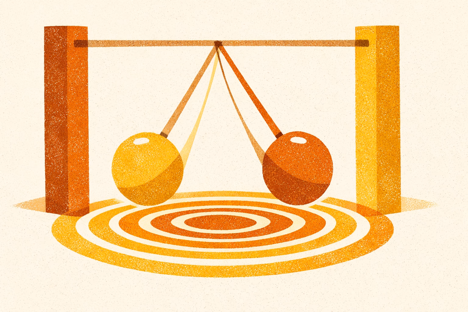
    The most widely accepted explanation involves a mechanism called central gain. When the tiny hair cells in the cochlea — the snail-shaped structure in the inner ear that converts sound waves into electrical signals — are damaged or lost, the amount of auditory input reaching the brain drops. The brain responds by effectively turning up its own internal volume, amplifying neural activity to compensate for the reduced input. This increased gain in the auditory pathway, at the cochlear nucleus, the inferior colliculus, and the auditory cortex, produces spontaneous electrical activity that the brain interprets as sound, even when none is present.

    A useful analogy: imagine turning up a stereo amplifier when the signal source has gone quiet. The amplifier starts reproducing the noise in its own circuits — a hiss or hum — because the gain is set too high for the level of input arriving. Your auditory system is doing something similar.

    The central gain model is supported by neuroscience research and appears to be the primary mechanism, though other pathways in the auditory cortex also contribute. For most people, the amplifier analogy captures the essential process accurately enough to be useful.

    Tinnitus is a phantom perception: a sound generated by the brain, not by any source in the environment. Over 99% of cases are subjective — only the person with tinnitus can hear it.

    How Common Is Tinnitus?

    If tinnitus feels isolating, the epidemiology tells a different story. A 2022 systematic review and meta-analysis of 113 studies — the most comprehensive analysis of global tinnitus prevalence conducted to date — found that approximately 14.4% of adults worldwide experience tinnitus, representing over 740 million people (Jarach et al. 2022). More than 120 million of those live with severe tinnitus. US estimates suggest more than 50 million Americans may be affected, though this figure derives from older survey data.

    Age is the strongest demographic predictor. Prevalence rises from around 9.7% in adults aged 18 to 44, to 13.7% in those aged 45 to 64, and reaches 23.6% in adults aged 65 and over (Jarach et al. 2022). The condition can and does occur at any age, including in children and young adults — often following noise exposure or ear infection.

    Contrary to older assumptions, the same large review found no significant difference in prevalence between men and women.

    It is worth separating transient tinnitus — the brief ringing after a loud noise or in a very quiet room, lasting seconds to minutes — from persistent tinnitus, which continues beyond a few days. Transient tinnitus is nearly universal and generally not a clinical concern. Chronic tinnitus, defined in Jarach et al. (2022) as lasting six months or longer, affects approximately 9.8% of adults globally. Tinnitus lasting three months or more — the threshold used in most clinical guidelines — encompasses a somewhat broader population.

    Why Tinnitus Happens: Causes and Risk Factors

    Tinnitus is a symptom, not a diagnosis in itself. In the majority of cases it reflects an underlying change in the auditory system, though in some people no specific cause is ever identified. Understanding the range of possible causes is the first step toward knowing what tests might help and whether a treatable condition is driving the sound.

    Auditory and cochlear causes

    Noise-induced hearing loss is the single most common cause of tinnitus. Prolonged or intense exposure to loud sound damages the cochlear hair cells described above — and once those cells are lost, they do not regenerate. Occupational noise (construction, manufacturing, music), recreational exposure (concerts, headphones at high volume), and single-event acoustic trauma (explosions, gunshots) all carry risk.

    Age-related hearing loss, known as presbycusis, follows a similar mechanism. As hair cell populations naturally decline with age, the central auditory system compensates with increased gain — which is one reason tinnitus becomes more common after the age of 60.

    The majority of people with tinnitus have some degree of co-occurring hearing loss, and many are unaware of it until formal testing. The exact figure varies across studies and clinical populations.

    Structural ear causes

    Several conditions affecting the structure of the ear can produce or contribute to tinnitus:

    • Earwax impaction: A blockage in the ear canal changes the acoustic environment and can cause or worsen tinnitus. This is one of the most easily treated causes.
    • Ear infections: Acute or chronic middle ear infections produce inflammation and fluid that can affect both hearing and tinnitus perception.
    • Ménière’s disease: A disorder of fluid pressure in the inner ear that typically causes episodic vertigo, fluctuating hearing loss, a sensation of fullness, and tinnitus — often described as a low-frequency roaring.
    • Otosclerosis: Abnormal bone growth in the middle ear that stiffens the ossicular chain and reduces sound transmission, leading to hearing loss and often tinnitus.

    Systemic and medical causes

    Several general health conditions are associated with tinnitus, likely through their effects on blood flow to the cochlea or on neural function:

    • Cardiovascular disease and hypertension
    • Diabetes
    • Thyroid disorders (both hypothyroidism and hyperthyroidism)
    • Anaemia

    Medications

    A number of medications are ototoxic — capable of damaging the inner ear — and can cause or worsen tinnitus as a side effect. These include certain aminoglycoside antibiotics (such as gentamicin), some chemotherapy agents (particularly cisplatin), high-dose aspirin, and some non-steroidal anti-inflammatory drugs (NSAIDs). If you notice tinnitus or a change in hearing after starting a new medication, let your prescribing doctor know. Do not stop a prescribed medication without speaking to your doctor first.

    If you develop tinnitus or changes in hearing after starting a new medication, tell your doctor promptly. Never stop a prescribed medication without medical advice.

    Head and neck causes

    The auditory system does not operate in isolation. Problems in the jaw, neck, and skull can influence tinnitus:

    • Temporomandibular joint (TMJ) disorder: The jaw joint sits close to the ear canal, and dysfunction there can produce clicking, ringing, or a sense of fullness in the ear.
    • Cervical spine problems: Neck injuries or degenerative changes can affect the neural and vascular supply to the auditory system.
    • Head trauma: Concussion and traumatic brain injury are associated with tinnitus, sometimes with delayed onset.

    Pulsatile tinnitus

    Pulsatile tinnitus — a rhythmic sound that beats in time with your pulse — is a distinct subtype that warrants separate mention and prompt medical evaluation. Unlike the steady-state phantom sounds of typical tinnitus, pulsatile tinnitus usually reflects an actual physical sound source, most commonly turbulent blood flow near the ear. Causes range from benign (such as increased awareness of normal blood flow) to conditions requiring treatment, including vascular malformations, high blood pressure, or rarely a tumour affecting blood vessels near the ear. Pulsatile tinnitus always warrants investigation.

    In many cases of tinnitus, no specific cause is ever found even after thorough investigation. This is not a failure of the diagnostic process — it reflects the fact that the neural changes underlying tinnitus often occur at a level too subtle to appear on standard imaging or hearing tests.

    Acute vs. Chronic Tinnitus: Does It Go Away?

    This is the question almost every person with new-onset tinnitus arrives with, and you deserve a direct, honest answer.

    Clinicians generally define acute tinnitus as lasting less than three months, and chronic tinnitus as persisting beyond three months (AWMF S3 guideline; NIDCD). The distinction matters because prognosis differs substantially between the two.

    What the evidence on remission actually shows

    You may have read that around 70% of acute tinnitus cases resolve spontaneously. This figure comes from studies of a specific population: people who developed tinnitus following idiopathic sudden sensorineural hearing loss (ISSNHL) — a type of sudden, significant hearing drop — with mild to moderate hearing impairment. In that group, Mühlmeier et al. (2016) found approximately two-thirds (around 65%) of patients had complete tinnitus remission at three months. The figure is real, but it applies to that specific context.

    For people who develop tinnitus in other circumstances — without significant sudden hearing loss, or in a general clinical setting — the prognosis is less clear-cut. A prospective study by Wallhäusser-Franke et al. (2017) followed 47 patients with tinnitus of four weeks or less and found that full remission had occurred in only 11% at six months. A companion review of similar studies noted remission rates consistently below 20% in general acute tinnitus clinic populations.

    What this means in practical terms: if your tinnitus appeared suddenly alongside significant hearing loss, there is meaningful evidence that it may resolve. If it arose in other circumstances, remission is less certain — but improvement is still possible, and early intervention improves outcomes.

    The widely cited ~70% remission figure applies to tinnitus following sudden sensorineural hearing loss. For acute tinnitus more broadly, many cases improve, but full remission is less certain. Early evaluation is important regardless.

    What happens if tinnitus becomes chronic

    For tinnitus that persists beyond three months, the goal shifts from hoping for resolution to achieving what clinicians call habituation. Habituation is the process by which the brain learns to deprioritise the tinnitus signal — to classify it as irrelevant background noise that no longer demands attention. This is not the same as the tinnitus disappearing; the sound may still be detectable if you listen for it. The difference is that it no longer triggers distress or disrupts functioning.

    Habituation is achievable for the majority of people with chronic tinnitus, particularly with structured support. The evidence-based therapies in the treatment section below are all aimed at supporting this process. Late spontaneous remission — tinnitus resolving after the chronic phase — does occur in some people, though no strong longitudinal data exists to quantify how often.

    The psychological state at acute onset also matters. Wallhäusser-Franke et al. (2017) found that high tinnitus distress and depression at the acute stage were predictors of a more difficult transition to chronic tinnitus — which is one reason early psychological support is genuinely valuable, not just a secondary consideration.

    “I kept waiting for the ringing to stop. When it didn’t, I thought that was it — that this was my life now, forever. What my audiologist helped me understand was that the goal wasn’t necessarily silence. It was getting to a place where the sound stopped running my day. That shift changed everything.”

    — Patient account shared through the American Tinnitus Association

    How Tinnitus Affects Daily Life

    Tinnitus loudness and the suffering it causes do not move in lockstep. Someone with relatively quiet tinnitus can be severely affected, while another person with objectively louder tinnitus copes well. This disconnect is real and well-documented — and it means that dismissive responses like “but it’s only quiet” entirely miss the point.

    A cross-sectional study of 163 adults with tinnitus (Musleh et al. 2024) provides a clear picture of the functional and emotional burden. Among those assessed:

    • 38.0% reported fatigue
    • 37.4% reported concentration difficulties
    • 36.8% reported sleep disturbances
    • 33.7% reported interference with daily activities
    • 30.1% reported reduced social participation

    The emotional impact was equally significant: 47.9% reported anger, 43.6% reported anxiety, 36.8% reported desperation, 30.7% reported depression.

    According to the American Tinnitus Association’s patient education materials (2018), between 48% and 78% of people with severe tinnitus experience a comorbid behavioural disorder — depression, anxiety, or another condition. These are not minor secondary effects.

    The feedback loop that makes things worse

    Tinnitus distress is not simply proportional to the volume of the sound. There is a psychological feedback loop at work: anxiety about tinnitus increases the amount of attention the brain directs toward it, which makes the sound more salient, which increases anxiety. Over time, this loop can amplify distress well beyond what the underlying sound would warrant.

    Clinicians distinguish between compensated tinnitus — where the sound is present but does not significantly disrupt daily functioning — and decompensated tinnitus, where distress and functional impairment are substantial. The same person can move between these states depending on life circumstances, stress levels, and whether they have access to effective support.

    CBT, the treatment with the strongest evidence base for tinnitus, works precisely by interrupting this feedback loop — changing the cognitive and emotional response to tinnitus rather than eliminating the sound itself.

    Getting Diagnosed: What to Expect

    If your tinnitus is new, persistent, or bothering you, a medical evaluation is the right first step. Understanding the tinnitus diagnosis process helps you know what to expect and what each test is looking for.

    Step one: your GP or primary care doctor

    Your first appointment will usually involve a detailed history — when the tinnitus started, what it sounds like, whether it is in one ear or both, whether hearing has changed, and whether there are any associated symptoms such as vertigo or ear pain. The doctor will examine your ear canals with an otoscope to check for visible causes like earwax impaction or infection, and may perform a brief hearing check.

    Many cases are referred from this point for specialist assessment.

    Step two: ENT or audiology referral

    An ear, nose and throat (ENT) specialist or audiologist will conduct more detailed testing. A pure-tone audiogram maps hearing thresholds across a range of frequencies and will usually identify any hearing loss that co-occurs with tinnitus. Tympanometry assesses how the eardrum and middle ear are functioning. These tests are painless and typically take 30 to 60 minutes.

    Validated questionnaires — such as the Tinnitus Handicap Inventory (THI) — are used to measure how much tinnitus is affecting daily life and to track whether treatment is helping over time (Musleh et al. 2024).

    Step three: imaging

    Not everyone with tinnitus needs a scan. The AAO-HNS guideline and clinical consensus indicate that imaging is warranted when tinnitus is:

    • Unilateral (one ear only)
    • Pulsatile
    • Associated with asymmetric hearing loss or neurological symptoms

    In these situations, MRI or CT scanning is used to rule out structural causes, including vestibular schwannoma (a benign tumour on the hearing nerve) in the case of unilateral tinnitus.

    For bilateral, non-pulsatile tinnitus without neurological signs, imaging is generally not required.

    When tests come back normal

    For many people, the audiogram and physical examination return results within normal limits or show only mild hearing loss, with no structural cause identified. This can feel frustrating when you are searching for an explanation. In practice, it is a meaningful finding: it means there is no serious underlying condition driving the tinnitus, and it focuses attention on the management strategies that are most likely to help.

    NICE guidelines (NICE 2020) recommend that information and tinnitus support be offered at all stages of care, not just after a cause is found.

    Treatment Options That Work

    Understanding tinnitus causes and treatment options together helps clarify why certain therapies work better than others. No treatment currently eliminates tinnitus reliably. What the evidence does support — clearly, across multiple well-designed trials — is that the distress and disruption caused by tinnitus can be significantly reduced. That is not a consolation prize. For most people, it is the outcome that matters most.

    The treatments below are presented in order of their evidence strength, not their popularity.

    1. Cognitive behavioural therapy (CBT) — strongest evidence

    CBT is the most evidence-supported treatment for tinnitus distress. A Cochrane systematic review (Fuller et al. 2020) analysed 28 randomised controlled trials involving 2,733 participants — all with tinnitus lasting three months or more. CBT reduced tinnitus-related distress with a standardised mean difference of -0.56 compared to no intervention (equivalent to approximately 10.9 points lower on the 100-point Tinnitus Handicap Inventory, exceeding the minimum clinically important difference of 7 points). The effect was maintained at follow-up. CBT also showed moderate-certainty evidence of benefit compared to audiological care alone.

    In a network meta-analysis of 22 RCTs (Lu et al. 2024), CBT ranked most effective for tinnitus-related distress outcomes. The AAO-HNS guideline gives CBT its highest recommendation level.

    How it works: CBT does not reduce the loudness of tinnitus. It changes the cognitive and emotional response to the sound — reducing the anxiety and hypervigilance that amplify distress and teaching the brain to deprioritise the tinnitus signal. Most CBT programmes for tinnitus run over 6 to 12 weeks and can be delivered in-person, in groups, or via digital platforms. CBT requires active participation and is not a passive treatment.

    2. Hearing aids — highly effective for the majority

    Because the majority of people with tinnitus have some degree of co-occurring hearing loss, hearing aids are relevant for a large proportion of those affected. Restoring auditory input through amplification directly addresses the central gain mechanism driving tinnitus — when the brain receives more sound from the environment, the compensatory overactivity that produces phantom sound tends to reduce. Multiple systematic reviews, including Chen et al. (2025), confirm consistent benefit from hearing aids in this population.

    Speaking with an audiologist about your hearing is a practical, low-risk early step.

    3. Tinnitus retraining therapy (TRT) — widely used, clinically valuable

    TRT combines structured counselling (based on the Jastreboff neurophysiological model of tinnitus) with low-level sound enrichment — typically a broadband noise generator worn in the ear. The aim is to facilitate habituation: training the brain over time to classify tinnitus as a neutral, irrelevant signal.

    TRT is widely used and guideline-endorsed. Its evidence base is less comprehensive than CBT’s in terms of randomised controlled trials — the Cochrane CBT review identified only one head-to-head comparison (n=42), which favoured CBT. An RCT (Luyten et al. 2020) compared TRT combined with EMDR versus TRT combined with CBT, finding clinically meaningful improvement in both arms (mean TFI decrease of 15.1 points in the TRT+CBT group, above the 13-point clinical significance threshold) with no statistically significant difference between them. TRT and CBT target overlapping mechanisms through different approaches, and some clinics offer both in combination.

    4. Sound therapy and masking

    Sound therapy covers a range of approaches that use external sound to reduce the perceptual contrast between tinnitus and the acoustic environment. This includes white noise generators, wearable sound enrichment devices, and structured music-based approaches. The underlying logic is straightforward: tinnitus is often more noticeable in quiet environments, because the brain has less external input to process.

    A Cochrane review (Sereda et al. 2018) of 8 RCTs found no evidence of superiority over waiting list controls or placebo in controlled comparisons, though within-group improvements were observed. Sound therapy is considered optional support rather than a primary treatment, but it is low-risk and many people find it practically helpful, particularly for sleep.

    A network meta-analysis (Lu et al. 2024) ranked sound therapy most effective for tinnitus handicap specifically, suggesting it may have particular value for functional impairment even if its effect on distress is less clear.

    5. Acceptance and commitment therapy (ACT)

    ACT is a psychological approach that focuses on changing your relationship to difficult experiences — including tinnitus — rather than trying to eliminate or control them. In the network meta-analysis by Lu et al. (2024), ACT ranked most effective for insomnia outcomes in tinnitus patients, suggesting it may be particularly useful for those whose primary difficulty is sleep disruption related to tinnitus.

    6. Medications

    No medication is approved to treat tinnitus itself, and none has been shown to reliably reduce the perception of phantom sound. The AAO-HNS guideline recommends against prescribing antidepressants, anticonvulsants, or supplements (including ginkgo biloba) specifically for tinnitus. The VA/DoD 2024 clinical practice guideline concludes that no drug treatment, vitamin, or herbal supplement has been shown to be more effective than placebo for tinnitus.

    Medications may appropriately address secondary symptoms: melatonin can support sleep, and antidepressants or anxiolytics may be warranted when depression or anxiety is a comorbidity in its own right. These decisions should be made with your doctor based on your full clinical picture — not as a route to silencing tinnitus directly.

    No medication currently approved for tinnitus treatment reliably reduces the sound. Be cautious of any product claiming to cure or eliminate tinnitus — no such treatment has been validated in high-quality clinical trials.

    7. Lifestyle and self-management

    Several self-management strategies have good practical rationale, even if large RCTs are limited:

    • Sleep hygiene: Poor sleep and tinnitus interact in both directions — tinnitus disrupts sleep, and sleep deprivation makes tinnitus more distressing. Structured sleep approaches (consistent sleep and wake times, reducing screen use before bed, quiet background sound) address both problems.
    • Stress management: Tinnitus distress typically increases during periods of high stress. Approaches that reduce overall arousal — exercise, relaxation techniques, mindfulness — can reduce the emotional salience of tinnitus.
    • Noise protection: Continued loud noise exposure accelerates the cochlear damage that drives tinnitus and worsens prognosis. Hearing protection in noisy environments is important both for tinnitus management and general hearing health.
    • Caffeine and alcohol: Patient reports of worsening tinnitus after caffeine and alcohol are common, though clinical trial evidence is limited. Individual responses vary; it is reasonable to experiment and observe.

    When to Act Fast: Red Flags That Need Immediate Attention

    Knowing when to see a doctor for tinnitus — and how urgently — can make a real difference to outcomes. The great majority of tinnitus cases do not represent a medical emergency, but some presentations require prompt evaluation — not routine GP booking, but same-day or urgent contact with a healthcare provider.

    Sudden hearing loss alongside tinnitus

    If tinnitus has appeared at the same time as a significant drop in hearing — particularly if this happened suddenly over hours or days — seek same-day or next-day evaluation. The treatment window for sudden sensorineural hearing loss can be as short as 72 hours. NICE guidelines (NICE 2020) recommend referral within 24 hours for sudden hearing loss that developed over 3 days or less and occurred within the last 30 days. Early treatment significantly improves the chance of hearing recovery and may also affect tinnitus outcomes.

    Tinnitus in one ear only

    Unilateral tinnitus — affecting only one ear, especially if persistent — warrants imaging to rule out vestibular schwannoma (also called an acoustic neuroma), a benign but significant tumour on the hearing nerve. Most cases turn out to have a more straightforward explanation, but unilateral tinnitus should not be left uninvestigated.

    Pulsatile tinnitus

    A rhythmic sound that pulses with your heartbeat always requires vascular investigation. Most causes are benign, but pulsatile tinnitus can occasionally indicate conditions affecting blood vessels near the ear that benefit from early identification.

    Tinnitus with vertigo, dizziness, or neurological symptoms

    Tinnitus accompanied by severe vertigo, facial weakness, sudden vision changes, or other neurological symptoms may indicate central nervous system involvement and requires urgent evaluation — same-day in most guidelines.

    Sudden hearing loss accompanied by tinnitus: seek same-day evaluation. The treatment window for sudden hearing loss can be as short as 72 hours. Do not wait for a routine appointment.

    These situations represent a minority of tinnitus cases overall. The point is not to alarm you — it is to make sure that the small number of presentations that need urgent attention get it promptly.

    Living With Tinnitus: What the Evidence Says About Outcomes

    You came to this page, most likely, because a sound arrived in your ears that you did not ask for and you needed to understand what it meant. The fear that comes with that — about permanence, about what it signals, about what life might look like if it stays — is a completely rational response to something genuinely disorienting.

    Here is what the evidence actually tells us. Tinnitus is common, affecting more than one in seven adults worldwide (Jarach et al. 2022). It rarely signals anything dangerous. For people whose tinnitus follows sudden hearing loss, there is meaningful evidence that resolution is possible in many cases — particularly with early evaluation and treatment. For those whose tinnitus persists, the outcomes are not simply “learn to live with it”: a Cochrane review of 28 RCTs demonstrates that CBT significantly reduces tinnitus-related distress, and hearing aids, TRT, and sound therapy add further tools to what is now a well-developed area of specialist care (Fuller et al. 2020; Chen et al. 2025).

    Habituation — the brain learning to deprioritise tinnitus as irrelevant background — is achievable for most people with chronic tinnitus who engage with appropriate support. That is not the same as disappearance, but for many people it amounts to the same thing in practice: a sound that was once overwhelming becomes something that can be present without running the day.

    The American Tinnitus Association puts it directly: there are evidence-based treatments that can significantly reduce the effect of tinnitus on daily activities and improve quality of life (American Tinnitus Association 2018). No one needs to accept dismissal or silence in the face of a real, disruptive symptom.

    This guide is the starting point. The satellite articles on this site go deeper into specific topics: the full evidence base for CBT, managing tinnitus-related sleep disruption, what the research pipeline looks like, and how to separate evidence-based supplements from those that do not hold up to scrutiny. Whatever the next question is, you do not have to work through it alone.

  • Tinnitus Research Digest: Treatment-Resistant Inner Ear Disease, Music Perception, and Brain Plasticity

    This week’s digest covers four items across tinnitus and inner ear research: a new Chinese clinical consensus on conditions that resist standard treatment, a study on music perception difficulties in tinnitus patients with normal audiograms, an older review of the brain changes thought to drive tinnitus, and a preclinical study on how the basal ganglia may affect sound filtering. The items range from clinically applicable to basic science with no immediate treatment implications.

  • Tinnitus Research Digest: ICBT Long-Term Data, Digital CBT, Musical Ear Syndrome, and Vestibular Schwannoma

    This week’s digest covers four studies relevant to people living with tinnitus and related auditory conditions. The items range from a six-year follow-up of internet-based CBT — one of the longest tinnitus therapy outcome studies to date — to a case report on musical hallucinations in a young adult, a clinical review of digital CBT, and a comparative radiotherapy study for vestibular schwannoma patients managing tinnitus alongside tumour treatment.

  • Tinnitus Research Digest: CBT Durability, Brainstem Findings, and Cardiovascular Links

    This week’s digest covers five studies spread across basic science, diagnostics, and management. The clearest take for patients comes from a six-year follow-up of internet-based CBT, which shows treatment benefits can last well beyond the initial programme. Two neurophysiology studies examine how the brain and brainstem behave in tinnitus — findings that deepen understanding without yet changing treatment. A large population study adds to the evidence linking tinnitus to cardiovascular conditions, and a small pilot tests an integrated care model worth watching.

  • Tinnitus Research Digest: Imaging, Mental Health, Physical Therapy, and Treatment Studies

    This week’s digest covers five studies spanning the biological, psychological, and physical dimensions of tinnitus. One imaging study offers insight into why a specific subtype of pulsatile tinnitus worsens over time. A cross-sectional study reinforces the scale of depression and anxiety in tinnitus clinic populations. Research on somatosensory tinnitus maps the physical dysfunctions that may be treatable. A retrospective study tests a nerve block intervention, and a long-term radiotherapy comparison addresses outcomes for acoustic neuroma patients.

  • Tinnitus Research Digest: Low-Frequency Hums, Anxiety and the Brain, Cochlear Implant Sound Sensitivity, and Heart Disease Links

    This week’s digest covers five studies across different aspects of tinnitus research. The items range from a question many patients carry quietly — whether low-frequency humming is real — to how anxiety shapes brainstem responses, how cochlear implant users experience sound sensitivity, and what a large population study tells us about tinnitus and heart disease. One older preclinical study rounds out the set with mechanistic context.

  • Tinnitus Research Digest: Mental Health Links and Early-Stage Brain Research

    This week’s digest covers two areas of tinnitus research: the well-documented overlap between tinnitus and mental health conditions, and early-stage work on objective measurement tools and brain-based biomarkers. The mental health review has the most direct relevance for patients managing tinnitus day to day. The remaining items reflect ongoing basic and methodological research that has not yet produced clinical applications.

  • Tinnitus Research Digest: Two Trials Recruiting, Animal Study, and a Debate Over Definition

    This week’s digest covers five items spanning clinical trials, basic science, and foundational theory. Two ongoing randomised trials are recruiting patients — one testing internet-delivered CBT in Canada, one comparing vagus nerve stimulation combined with custom music therapy against music therapy alone. A preclinical study examines light-based therapy targeting overactive auditory brain circuits in animal models. A 2021 review of drug-induced tinnitus mechanisms rounds out the applied research. Finally, a philosophical paper asks whether tinnitus has ever been properly defined — a question with real consequences for how research is designed and measured.

Subscribe to Our Tinnitus Newsletter

  • Learn everything about tinnitus causes, myths, and treatments
  • Get the latest tinnitus research delivered to your inbox every week

You can unsubscribe anytime.